Multiple myeloma x ray: Difference between revisions
Jump to navigation
Jump to search
(→X Ray) |
|||
| Line 3: | Line 3: | ||
{{CMG}} | {{CMG}} | ||
==Overview== | ==Overview== | ||
On X-ray radiography, multiple myeloma is characterized by punched out bony lesions and hair on end appearance. | On X-ray radiography, multiple myeloma is characterized by punched out bony lesions, generalized osteopaenia, and hair on end appearance. | ||
==X Ray== | ==X Ray== | ||
Revision as of 19:50, 20 September 2015
|
Multiple myeloma Microchapters |
|
Diagnosis |
|---|
|
Treatment |
|
Case Studies |
|
Multiple myeloma x ray On the Web |
|
American Roentgen Ray Society Images of Multiple myeloma x ray |
|
Risk calculators and risk factors for Multiple myeloma x ray |
Editor-In-Chief: C. Michael Gibson, M.S., M.D. [1]
Overview
On X-ray radiography, multiple myeloma is characterized by punched out bony lesions, generalized osteopaenia, and hair on end appearance.
X Ray
- Simple radiography is the current gold standard for the initial diagnosis and evaluation of relapses of multiple myeloma. The long bones and the spine must always be evaluated while the evaluation of other bones merit consideration based on the symptoms of the patient.
- Shown below are images depicting, the involvement of skull and spinal cord respectively in a case of multiple myeloma.
-
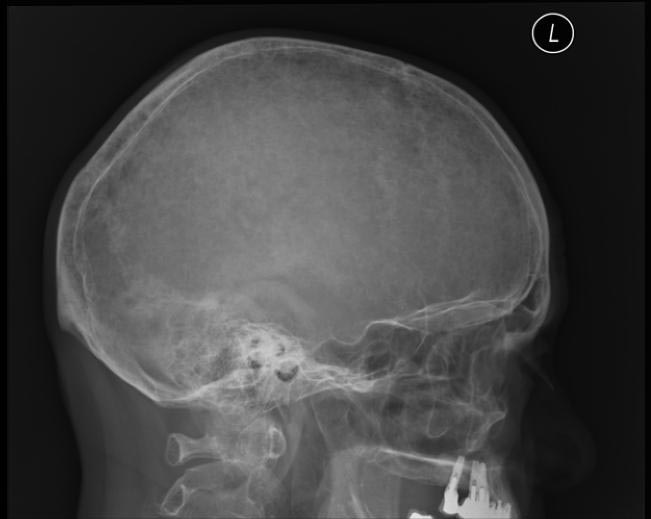
X ray showing hair on end appearance.
-
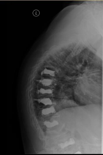
X ray spine showing collapsed vertebrae.
-
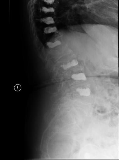
X ray spine showing increased space between 2 vertebrae suggestive of possible malignancy.


