Multiple myeloma x ray: Difference between revisions
Hannan Javed (talk | contribs) (→X-ray) |
Hannan Javed (talk | contribs) No edit summary |
||
| Line 4: | Line 4: | ||
==Overview== | ==Overview== | ||
X-ray may be helpful in the diagnosis of multiple myeloma. Findings on X-ray suggestive of multiple myeloma include punched out bony lesions, generalized [[osteopenia]], and hair-on-end appearance.<ref name="radio">Multiple myeloma. Radiopaedia (2015)http://radiopaedia.org/articles/multiple-myeloma-1 Accessed on September, 20th 2015</ref><ref name="pmid24614435">{{cite journal |vauthors=Reisenbuckler C |title=Multiple myeloma and diagnostic imaging |journal=Radiol Technol |volume=85 |issue=4 |pages=391–410; quiz 411–3 |date=2014 |pmid=24614435 |doi= |url=}}</ref> In some cases, MRI may be needed if X-ray is insufficient for diagnosis. | [[X-ray]] may be helpful in the [[diagnosis]] of [[multiple myeloma]]. Findings on [[X-ray]] suggestive of [[multiple myeloma]] include punched out bony [[lesions]], generalized [[osteopenia]], and hair-on-end appearance.<ref name="radio">Multiple myeloma. Radiopaedia (2015)http://radiopaedia.org/articles/multiple-myeloma-1 Accessed on September, 20th 2015</ref><ref name="pmid24614435">{{cite journal |vauthors=Reisenbuckler C |title=Multiple myeloma and diagnostic imaging |journal=Radiol Technol |volume=85 |issue=4 |pages=391–410; quiz 411–3 |date=2014 |pmid=24614435 |doi= |url=}}</ref> In some cases, [[MRI]] may be needed if [[X-ray]] is insufficient for [[diagnosis]]. | ||
==X-ray== | ==X-ray== | ||
*Simple radiography is the current gold standard for the initial diagnosis and evaluation of lytic lesions of multiple myeloma.<ref name="radio">Multiple myeloma. Radiopaedia (2015)http://radiopaedia.org/articles/multiple-myeloma-1 Accessed on September, 20th 2015</ref> | *Simple [[radiography]] is the current [[Gold standard (test)|gold standard]] for the initial [[diagnosis]] and evaluation of lytic [[lesions]] of [[multiple myeloma]].<ref name="radio">Multiple myeloma. Radiopaedia (2015)http://radiopaedia.org/articles/multiple-myeloma-1 Accessed on September, 20th 2015</ref> | ||
*The [[long bone]]s and the [[spine]] must always be evaluated while the evaluation of other bones merit consideration based on the patient's symptoms.<ref name="radio">Multiple myeloma. Radiopaedia (2015)http://radiopaedia.org/articles/multiple-myeloma-1 Accessed on September, 20th 2015</ref> | *The [[long bone]]s and the [[spine]] must always be evaluated while the evaluation of other [[bones]] merit consideration based on the [[patient]]'s symptoms.<ref name="radio">Multiple myeloma. Radiopaedia (2015)http://radiopaedia.org/articles/multiple-myeloma-1 Accessed on September, 20th 2015</ref> | ||
*A series of plain films, or [[skeletal survey]], is essential in not only the diagnosis of multiple myeloma, but also in assessing response, and pre-empting potential complications (e.g. pathological fractures). A typical skeletal survey consists of the following films:<ref name="radio">Multiple myeloma. Radiopaedia (2015)http://radiopaedia.org/articles/multiple-myeloma-1 Accessed on September, 20th 2015</ref> | *A series of plain films, or [[skeletal survey]], is essential in not only the [[diagnosis]] of [[multiple myeloma]], but also in assessing response, and pre-empting potential [[complications]] (e.g. pathological [[Bone fracture|fractures]]). | ||
=== Skeletal survey === | |||
*A typical [[skeletal survey]] consists of the following films:<ref name="radio">Multiple myeloma. Radiopaedia (2015)http://radiopaedia.org/articles/multiple-myeloma-1 Accessed on September, 20th 2015</ref> | |||
:*Lateral [[skull]] | :*Lateral [[skull]] | ||
:*Frontal [[chest]] film | :*Frontal [[chest]] film | ||
| Line 16: | Line 19: | ||
:*[[Pelvis]] | :*[[Pelvis]] | ||
:*[[Femur]] | :*[[Femur]] | ||
:*[[Humerus]] [[File:Lytic lesion in right forearm.jpg|alt=Lytic lesions in multiple myeloma|Lytic lesions in multiple myeloma|center|frame]] | :*[[Humerus]] | ||
:*The vast majority of lesions seen on plain radiography are purely [[lytic]]. Lytic lesions are sharply defined and "punched-out" in appearance, with endosteal scalloping when abutting cortex. The lesions are sclerotic in only 3% of patients.<ref name="radio" /> [[File:Lytic lesion in right forearm.jpg|alt=Lytic lesions in multiple myeloma|Lytic lesions in multiple myeloma|center|frame]] | |||
*The vast majority of lesions seen on plain radiography are purely [[lytic]]. Lytic lesions are sharply defined and "punched-out" in appearance, with endosteal scalloping when abutting cortex. The lesions are sclerotic in only 3% of patients.<ref name="radio">Multiple myeloma. Radiopaedia (2015)http://radiopaedia.org/articles/multiple-myeloma-1 Accessed on September, 20th 2015</ref> | *The vast majority of lesions seen on plain radiography are purely [[lytic]]. Lytic lesions are sharply defined and "punched-out" in appearance, with endosteal scalloping when abutting cortex. The lesions are sclerotic in only 3% of patients.<ref name="radio">Multiple myeloma. Radiopaedia (2015)http://radiopaedia.org/articles/multiple-myeloma-1 Accessed on September, 20th 2015</ref> | ||
*The X-ray is the most inexpensive diagnostic modality for lytic lesions. In some cases, MRI may be warranted, as MRI has a higher sensitivity than X-ray. | *The X-ray is the most inexpensive diagnostic modality for lytic lesions. In some cases, MRI may be warranted, as MRI has a higher sensitivity than X-ray. | ||
Revision as of 15:06, 15 November 2018
|
Multiple myeloma Microchapters |
|
Diagnosis |
|---|
|
Treatment |
|
Case Studies |
|
Multiple myeloma x ray On the Web |
|
American Roentgen Ray Society Images of Multiple myeloma x ray |
|
Risk calculators and risk factors for Multiple myeloma x ray |
Editor-In-Chief: C. Michael Gibson, M.S., M.D. [1] Associate Editor(s)-in-Chief: Haytham Allaham, M.D. [2] Shyam Patel [3]
Overview
X-ray may be helpful in the diagnosis of multiple myeloma. Findings on X-ray suggestive of multiple myeloma include punched out bony lesions, generalized osteopenia, and hair-on-end appearance.[1][2] In some cases, MRI may be needed if X-ray is insufficient for diagnosis.
X-ray
- Simple radiography is the current gold standard for the initial diagnosis and evaluation of lytic lesions of multiple myeloma.[1]
- The long bones and the spine must always be evaluated while the evaluation of other bones merit consideration based on the patient's symptoms.[1]
- A series of plain films, or skeletal survey, is essential in not only the diagnosis of multiple myeloma, but also in assessing response, and pre-empting potential complications (e.g. pathological fractures).
Skeletal survey
- A typical skeletal survey consists of the following films:[1]
- Lateral skull
- Frontal chest film
- Cervico-thoraco-lumbar spine
- Shoulder
- Pelvis
- Femur
- Humerus
- The vast majority of lesions seen on plain radiography are purely lytic. Lytic lesions are sharply defined and "punched-out" in appearance, with endosteal scalloping when abutting cortex. The lesions are sclerotic in only 3% of patients.[1]

Lytic lesions in multiple myeloma

- The vast majority of lesions seen on plain radiography are purely lytic. Lytic lesions are sharply defined and "punched-out" in appearance, with endosteal scalloping when abutting cortex. The lesions are sclerotic in only 3% of patients.[1]
- The X-ray is the most inexpensive diagnostic modality for lytic lesions. In some cases, MRI may be warranted, as MRI has a higher sensitivity than X-ray.
- X-rays should be avoided in pregnant patients with multiple myeloma. MRI can be done instead.
- Shown below are images depicting the involvement of skull and spinal cord respectively in a case of multiple myeloma.
-
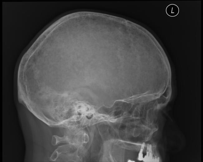
X ray showing hair on end appearance.
-
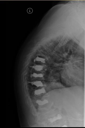
X ray spine showing collapsed vertebrae.
-
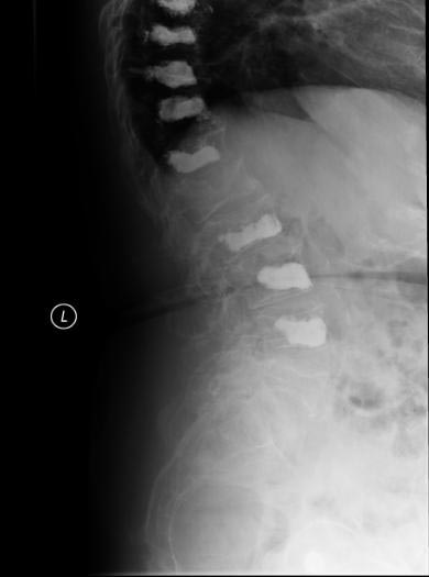
X ray spine showing increased space between 2 vertebrae suggestive of possible malignancy.


