Multiple myeloma MRI: Difference between revisions
Jump to navigation
Jump to search
Rim Halaby (talk | contribs) (→MRI) |
|||
| Line 3: | Line 3: | ||
{{CMG}} | {{CMG}} | ||
==Overview== | ==Overview== | ||
The workup of suspected multiple myeloma includes a [[skeletal survey]]. | The workup of suspected multiple myeloma includes a [[skeletal survey]].[[Magnetic resonance imaging]] (MRI) is more sensitive than simple X-ray in the detection of lytic lesions, and may supersede skeletal survey, especially when vertebral disease is suspected. | ||
==MRI== | ==MRI== | ||
Revision as of 01:05, 18 February 2014
|
Multiple myeloma Microchapters |
|
Diagnosis |
|---|
|
Treatment |
|
Case Studies |
|
Multiple myeloma MRI On the Web |
|
American Roentgen Ray Society Images of Multiple myeloma MRI |
Editor-In-Chief: C. Michael Gibson, M.S., M.D. [1]
Overview
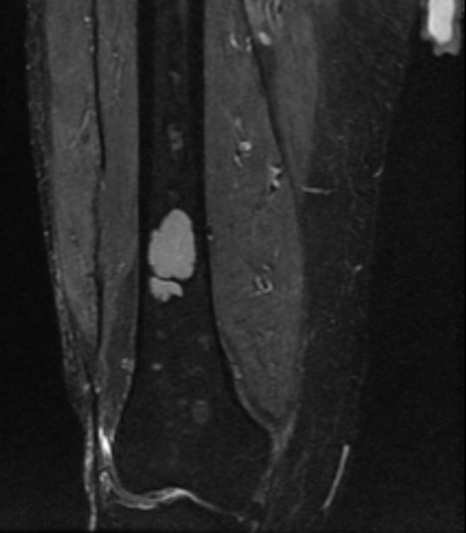
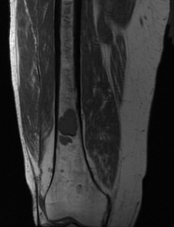
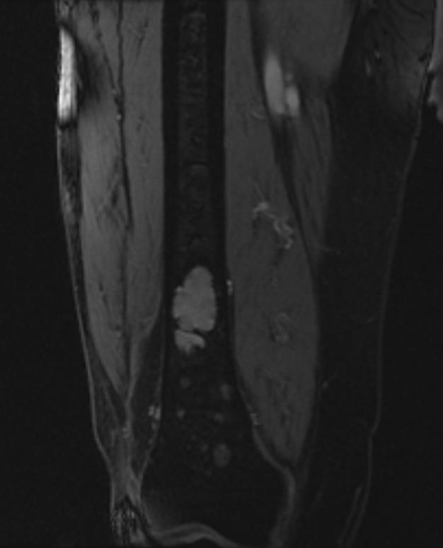
The workup of suspected multiple myeloma includes a skeletal survey.Magnetic resonance imaging (MRI) is more sensitive than simple X-ray in the detection of lytic lesions, and may supersede skeletal survey, especially when vertebral disease is suspected.
MRI
Shown below is a series of MRI images of long bones involved in multiple myeloma. (Images courtesy of RadsWiki)
-
Multiple myeloma
-
Multiple myeloma
-
Multiple myeloma


Shown below is a series of MRI images in a multiple myeloma patient complaining of back pain.

