Lung abscess CT: Difference between revisions
Jump to navigation
Jump to search
No edit summary |
Aditya Ganti (talk | contribs) |
||
| Line 10: | Line 10: | ||
<gallery> | <gallery> | ||
Image: | Image: | ||
*CT is helpful in differentiating the cavitation of abscess that cannot be clearly delineated on the chest radiograph from empyema and other cavitary lesions.<ref name="pmid6602513">{{cite journal |vauthors=Stark DD, Federle MP, Goodman PC, Podrasky AE, Webb WR |title=Differentiating lung abscess and empyema: radiography and computed tomography |journal=AJR Am J Roentgenol |volume=141 |issue=1 |pages=163–7 |year=1983 |pmid=6602513 |doi=10.2214/ajr.141.1.163 |url=}}</ref> | |||
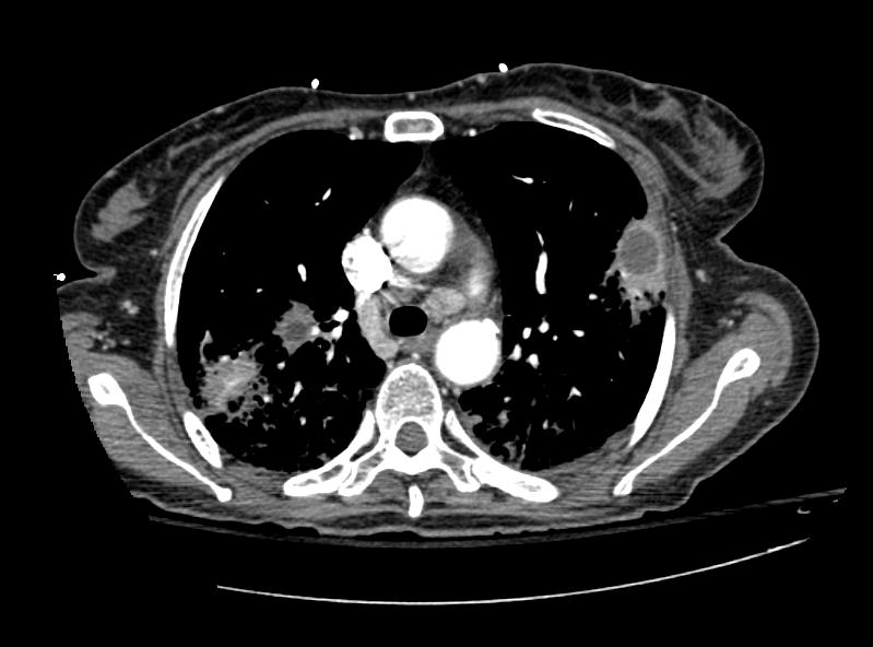
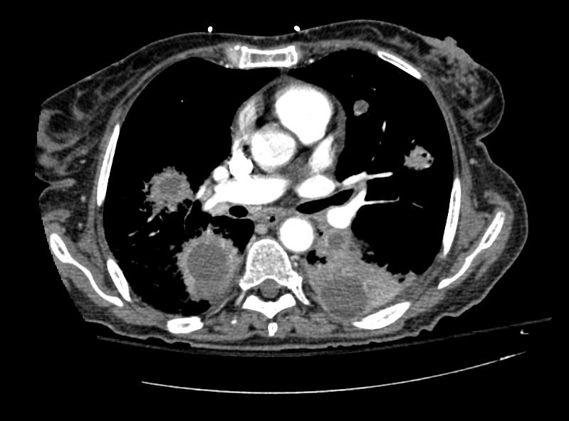
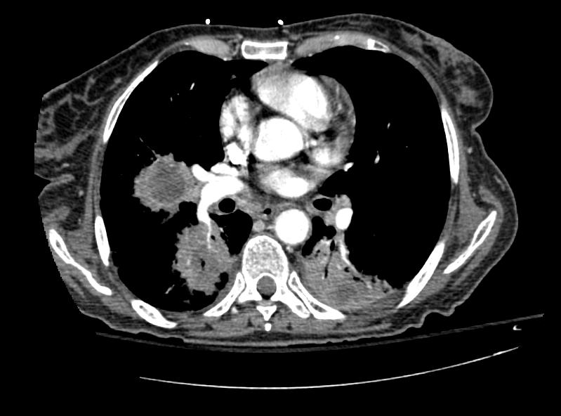
*On CT scan lung abscess is visualized as a rounded radiolucent lesion with a thick wall and ill-defined irregular margins, and is located within the parenchyma compared with loculated empyema, which may be difficult to distinguish on chest radiographs. <ref name="Mayer1982">{{cite journal|last1=Mayer|first1=Thom|title=Computed Tomographic Findings of Neonatal Lung Abscess|journal=Archives of Pediatrics & Adolescent Medicine|volume=136|issue=1|year=1982|pages=39|issn=1072-4710|doi=10.1001/archpedi.1982.03970370041010}}</ref> | |||
*Computed tomography (CT) lung is considered as the gold standard not only for the diagnosis of lung abscess but also for guiding therapeutic procedures such as trans-thoracic drainage of localized lung abscess .<ref name="BouhemadZhang2007">{{cite journal|last1=Bouhemad|first1=Bélaïd|last2=Zhang|first2=Mao|last3=Lu|first3=Qin|last4=Rouby|first4=Jean-Jacques|journal=Critical Care|volume=11|issue=1|year=2007|pages=205|issn=13648535|doi=10.1186/cc5668}}</ref> | |||
*CT scan is very helpful in excluding endobronchial obstruction due to malignancy or foreign body and provides additional information about size and location of the abscess, | |||
Pulmonary abscesses CT 101.jpg | Pulmonary abscesses CT 101.jpg | ||
Revision as of 14:00, 6 February 2017
|
Lung abscess Microchapters |
|
Diagnosis |
|
Treatment |
|
Case Studies |
|
Lung abscess CT On the Web |
|
American Roentgen Ray Society Images of Lung abscess CT |
Editor-In-Chief: C. Michael Gibson, M.S., M.D. [1]
Please help WikiDoc by adding more content here. It's easy! Click here to learn about editing.


