Liposclerosing myxofibrous tumor

Jump to navigation Jump to search
The printable version is no longer supported and may have rendering errors. Please update your browser bookmarks and please use the default browser print function instead.

WikiDoc Resources for Liposclerosing myxofibrous tumor

Articles

Most recent articles on Liposclerosing myxofibrous tumor

Most cited articles on Liposclerosing myxofibrous tumor

Review articles on Liposclerosing myxofibrous tumor

Articles on Liposclerosing myxofibrous tumor in N Eng J Med, Lancet, BMJ

Media

Powerpoint slides on Liposclerosing myxofibrous tumor

Images of Liposclerosing myxofibrous tumor

Photos of Liposclerosing myxofibrous tumor

Podcasts & MP3s on Liposclerosing myxofibrous tumor

Videos on Liposclerosing myxofibrous tumor

Evidence Based Medicine

Cochrane Collaboration on Liposclerosing myxofibrous tumor

Bandolier on Liposclerosing myxofibrous tumor

TRIP on Liposclerosing myxofibrous tumor

Clinical Trials

Ongoing Trials on Liposclerosing myxofibrous tumor at Clinical Trials.gov

Trial results on Liposclerosing myxofibrous tumor

Clinical Trials on Liposclerosing myxofibrous tumor at Google

Guidelines / Policies / Govt

US National Guidelines Clearinghouse on Liposclerosing myxofibrous tumor

NICE Guidance on Liposclerosing myxofibrous tumor

NHS PRODIGY Guidance

FDA on Liposclerosing myxofibrous tumor

CDC on Liposclerosing myxofibrous tumor

Books

Books on Liposclerosing myxofibrous tumor

News

Liposclerosing myxofibrous tumor in the news

Be alerted to news on Liposclerosing myxofibrous tumor

News trends on Liposclerosing myxofibrous tumor

Commentary

Blogs on Liposclerosing myxofibrous tumor

Definitions

Definitions of Liposclerosing myxofibrous tumor

Patient Resources / Community

Patient resources on Liposclerosing myxofibrous tumor

Discussion groups on Liposclerosing myxofibrous tumor

Patient Handouts on Liposclerosing myxofibrous tumor

Directions to Hospitals Treating Liposclerosing myxofibrous tumor

Risk calculators and risk factors for Liposclerosing myxofibrous tumor

Healthcare Provider Resources

Symptoms of Liposclerosing myxofibrous tumor

Causes & Risk Factors for Liposclerosing myxofibrous tumor

Diagnostic studies for Liposclerosing myxofibrous tumor

Treatment of Liposclerosing myxofibrous tumor

Continuing Medical Education (CME)

CME Programs on Liposclerosing myxofibrous tumor

International

Liposclerosing myxofibrous tumor en Espanol

Liposclerosing myxofibrous tumor en Francais

Business

Liposclerosing myxofibrous tumor in the Marketplace

Patents on Liposclerosing myxofibrous tumor

Experimental / Informatics

List of terms related to Liposclerosing myxofibrous tumor

Editor-In-Chief: C. Michael Gibson, M.S., M.D. [1]

Overview

Liposclerosing myxofibrous tumor (LSMFT) of bone, a benign fibro-osseous lesion, is characterized by a complex mixture of histologic elements. These include lipoma, fibroxanthoma, myxoma, myxofibroma, fibrous dysplasia –like features, cyst formation, fat necrosis, ischemic ossification, and, rarely, cartilage. LSMFT has a striking predilection for the femur (approximately 85% of the lesions). Approximately 91% of the femoral lesions are in the intertrochanteric region. The prevalence of malignant transformation in LSMFT has been reported to be 10%–16%. The increased propensity of LSMFT for malignant transformation is likely to be secondary to its extensive involutional and ischemic change, with the associated sarcoma arising from areas of ischemic ossification within the lesion or from progressive in situ atypism of the altered lipomatous elements.

Diagnosis

The imaging findings are

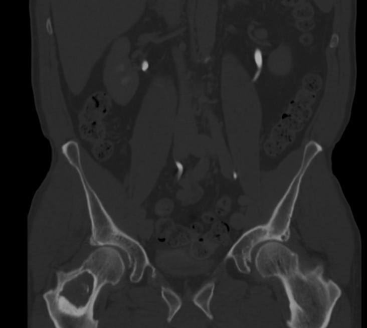
  • Radiographs typically showed a geographic lesion with a well-defined, often extensively sclerotic margin; such findings indicate an indolent pattern of growth.
  • Despite its name, lipomatous tissue was not identified on CT and MR imaging studies.
  • Myxoid tissue causes decreased attenuation seen in portions of the lesions on the CT scans and causes the high signal intensity seen on the T2-weighted MR images.
  • LSMFT can be readily distinguished from intraosseous lipoma on CT scans or MR images by the identification of fat within a lipoma.

CT images of a liposclerosing myxofibrous tumor in the right femur
References

Template:WH Template:WS