Hyperosmolar hyperglycemic state CT scan: Difference between revisions
No edit summary |
|||
| (5 intermediate revisions by 3 users not shown) | |||
| Line 1: | Line 1: | ||
__NOTOC__ | __NOTOC__ | ||
{{Hyperosmolar hyperglycemic state}} | {{Hyperosmolar hyperglycemic state}} | ||
{{CMG}}; {{AE}} | {{CMG}}; {{AE}} {{HS}} | ||
==Overview== | ==Overview== | ||
| Line 8: | Line 8: | ||
==CT scan== | ==CT scan== | ||
* CT scan may be helpful in the workup of hyperosmolar hyperglycemic state when a patient presents with an [[altered state of consciousness]] or with [[Focal neurologic signs|focal neurological signs and symptoms]] to rule out [[stroke]]. | * CT scan may be helpful in the workup of hyperosmolar hyperglycemic state when a patient presents with an [[altered state of consciousness]] or with [[Focal neurologic signs|focal neurological signs and symptoms]] to rule out [[stroke]]. | ||
* CT scan may also | |||
* The following findings can be seen in [[cerebral edema]] on CT scan: | * CT scan may also aid in the diagnosis of [[cerebral edema]] which can occur as a complication of treatment of hyperosmolar hyperglycemic state.<ref name="pmid3543638">{{cite journal |vauthors=Arieff AI |title=Cerebral edema complicating nonketotic hyperosmolar coma |journal=Miner Electrolyte Metab |volume=12 |issue=5-6 |pages=383–9 |year=1986 |pmid=3543638 |doi= |url=}}</ref> | ||
* The following findings can be seen in [[cerebral edema]] on CT scan:<ref name="pmid17521673">{{cite journal |vauthors=Park KY, Kim JH, Chung CS, Lee KH, Chung PW, Kim BM, Kim GM |title=Cortical sulcal effacement on brain CT associated with cerebral hyperperfusion after carotid artery stenting |journal=J. Neurol. Sci. |volume=260 |issue=1-2 |pages=83–6 |year=2007 |pmid=17521673 |doi=10.1016/j.jns.2007.04.010 |url=}}</ref><ref name="pmid17272776">{{cite journal |vauthors=Butcher KS, Lee SB, Parsons MW, Allport L, Fink J, Tress B, Donnan G, Davis SM |title=Differential prognosis of isolated cortical swelling and hypoattenuation on CT in acute stroke |journal=Stroke |volume=38 |issue=3 |pages=941–7 |year=2007 |pmid=17272776 |doi=10.1161/01.STR.0000258099.69995.b6 |url=}}</ref> | |||
** Diffuse effacement of the sulci and lateral ventricles | ** Diffuse effacement of the sulci and lateral ventricles | ||
** Hypoattenuation of the brain parenchyma | ** Hypoattenuation of the [[brain]] parenchyma | ||
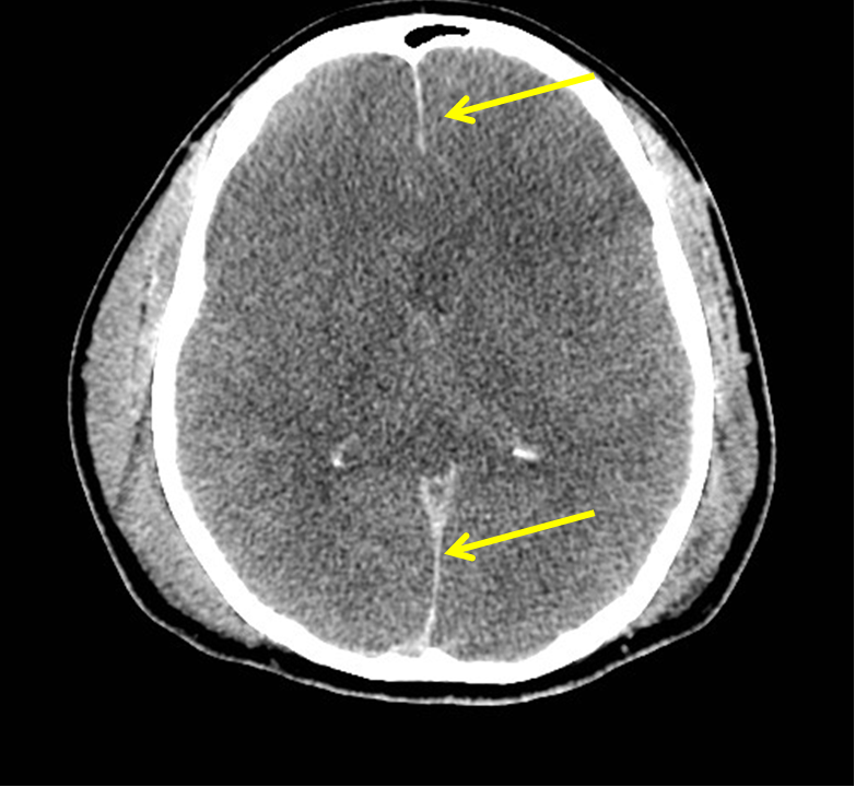
[[Image:Diffuse-cerebral-edema1_(1).png|400px|left|frame|'''Axial noncontrast CT scan of the head reveals diffuse effacement of the sulci and lateral ventricles and hypoattenuation of the brain parenchyma. Note also the falx appears hyperattenuating (yellow arrows)''', source: radiologypics.com]] | [[Image:Diffuse-cerebral-edema1_(1).png|400px|left|frame|'''Axial noncontrast CT scan of the head reveals diffuse effacement of the sulci and lateral ventricles and hypoattenuation of the brain parenchyma. Note also the falx appears hyperattenuating (yellow arrows)''', source: radiologypics.com]] | ||
<br style="clear:left"> | <br style="clear:left"> | ||
| Line 20: | Line 21: | ||
{{WH}} | {{WH}} | ||
{{WS}} | {{WS}} | ||
[[Category:Medicine]] | |||
[[Category:Endocrinology]] | |||
[[Category:Up-To-Date]] | |||
| |||
[[Category:Emergency medicine]] | |||
[[Category:Radiology]] | |||
Latest revision as of 17:46, 17 October 2017
|
Hyperosmolar hyperglycemic state Microchapters |
|
Differentiating Hyperosmolar hyperglycemic state from other Diseases |
|---|
|
Diagnosis |
|
Treatment |
|
Case Studies |
|
Hyperosmolar hyperglycemic state CT scan On the Web |
|
American Roentgen Ray Society Images of Hyperosmolar hyperglycemic state CT scan |
|
Risk calculators and risk factors for Hyperosmolar hyperglycemic state CT scan |
Editor-In-Chief: C. Michael Gibson, M.S., M.D. [1]; Associate Editor(s)-in-Chief: Husnain Shaukat, M.D [2]
Overview
CT scan may be helpful in the workup of hyperosmolar hyperglycemic state when a patient presents with an altered state of consciousness or with focal neurological signs and symptoms to rule out stroke. CT scan can also help in the diagnosis of cerebral edema which can occur as a complication of treatment of hyperosmolar hyperglycemic state.
CT scan
- CT scan may be helpful in the workup of hyperosmolar hyperglycemic state when a patient presents with an altered state of consciousness or with focal neurological signs and symptoms to rule out stroke.
- CT scan may also aid in the diagnosis of cerebral edema which can occur as a complication of treatment of hyperosmolar hyperglycemic state.[1]
- The following findings can be seen in cerebral edema on CT scan:[2][3]
- Diffuse effacement of the sulci and lateral ventricles
- Hypoattenuation of the brain parenchyma

References
- ↑ Arieff AI (1986). "Cerebral edema complicating nonketotic hyperosmolar coma". Miner Electrolyte Metab. 12 (5–6): 383–9. PMID 3543638.
- ↑ Park KY, Kim JH, Chung CS, Lee KH, Chung PW, Kim BM, Kim GM (2007). "Cortical sulcal effacement on brain CT associated with cerebral hyperperfusion after carotid artery stenting". J. Neurol. Sci. 260 (1–2): 83–6. doi:10.1016/j.jns.2007.04.010. PMID 17521673.
- ↑ Butcher KS, Lee SB, Parsons MW, Allport L, Fink J, Tress B, Donnan G, Davis SM (2007). "Differential prognosis of isolated cortical swelling and hypoattenuation on CT in acute stroke". Stroke. 38 (3): 941–7. doi:10.1161/01.STR.0000258099.69995.b6. PMID 17272776.