Herpes zoster physical examination: Difference between revisions
No edit summary |
No edit summary |
||
| Line 2: | Line 2: | ||
{{Herpes zoster}} | {{Herpes zoster}} | ||
{{CMG}}; L. Katie Morrison, MD; '''Associate Editor(s)-In-Chief:''' {{CZ}}; {{JH}}. | {{CMG}}; L. Katie Morrison, MD; '''Associate Editor(s)-In-Chief:''' {{CZ}}; {{JH}}. | ||
== Physical Examination == | == Physical Examination == | ||
| Line 9: | Line 7: | ||
The rash develops into clusters of clear vesicles. New vesicles continue to form over three to five days and progressively dry and crust over. They usually heal in two to four weeks. There may be permanent pigmentation changes and scarring on the skin. | The rash develops into clusters of clear vesicles. New vesicles continue to form over three to five days and progressively dry and crust over. They usually heal in two to four weeks. There may be permanent pigmentation changes and scarring on the skin. | ||
<gallery | ===Gallery=== | ||
=====Skin===== | |||
(Images shown below courtesy of Charlie Goldberg, M.D., UCSD School of Medicine and VA Medical Center, San Diego, CA) | |||
<gallery> | |||
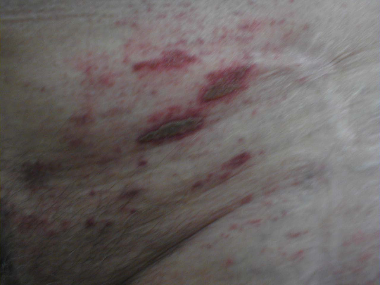
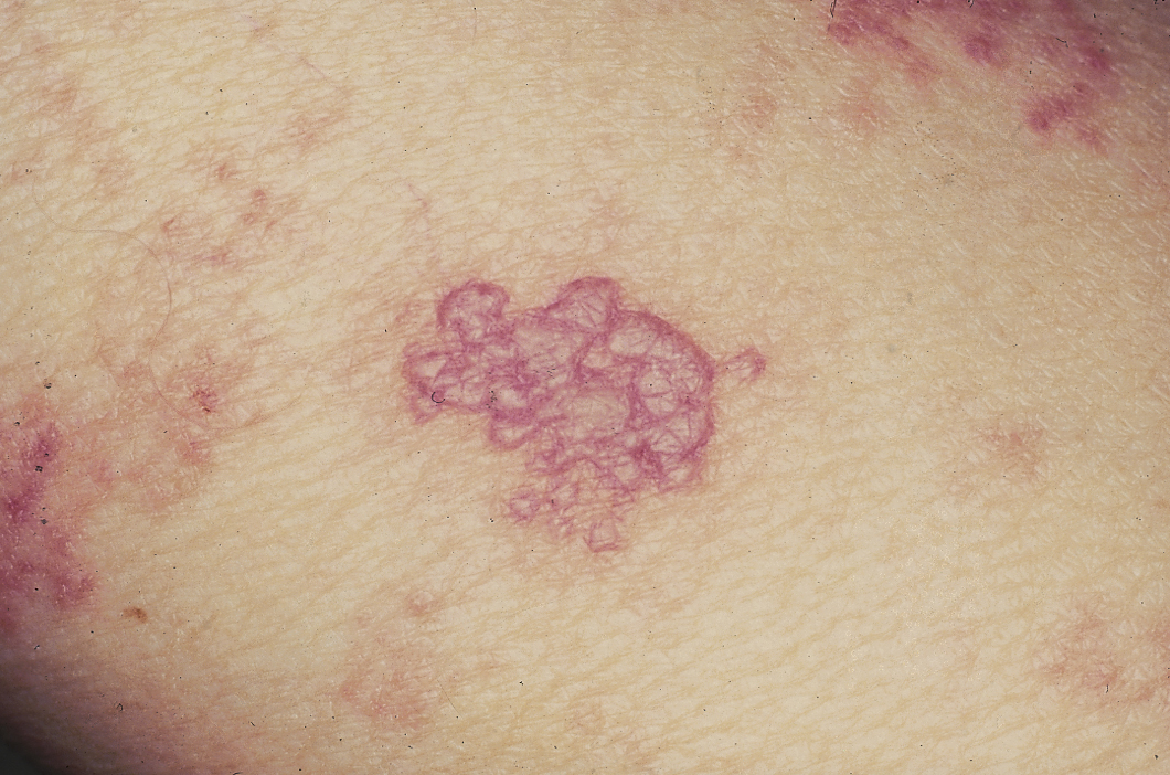
Image:Skin zoster buttocks1.jpg|Herpes Zoster: Coalesced [[vesicle]]s resulting from reactivation of HZV [[infection]]. | |||
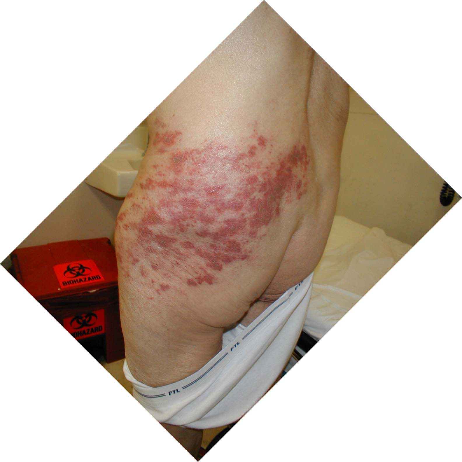
Image:Skin zoster buttocks2.jpg|Herpes Zoster: [[dermatome|Dermatomally]] distributed vesicles, many of which have coalesced, in patient with HZV infection. | |||
Image:Skin zoster buttocks4.jpg|Herpes Zoster: [[dermatome|Dermatomally]] distributed vesicles in patient with HZV infection. | |||
Image:Skin_zoster_buttocks3.jpg|Herpes Zoster: [[dermatome|Dermatomally]] distributed vesicles, many of which have coalesced, in patient with HZV infection. | |||
Image:Herpes zoster4.jpg|Shingles on waist | |||
Image:Herpes zoster1.jpg|[[dermatome|Dermatomal]] involvement of [[rash]] | Image:Herpes zoster1.jpg|[[dermatome|Dermatomal]] involvement of [[rash]] | ||
Image:Herpes zoster2.jpg|[[dermatome|Dermatomal]] involvement of skin rash | Image:Herpes zoster2.jpg|[[dermatome|Dermatomal]] involvement of skin rash | ||
| Line 17: | Line 27: | ||
Image:varicella zoster.jpg|Varicella zoster | Image:varicella zoster.jpg|Varicella zoster | ||
</gallery> | </gallery> | ||
<gallery> | |||
Image:skin_herpes_zoster.jpg|Herpes Zoster. <br> (Courtesy of Josh Fierer, M.D. and Charlie Goldberg, M.D.) | |||
Image:c3zoster.jpg|Herpes Zoster C3 Distribution: [[dermatome|Dermatomally]] distributed vesicles, many of which have coalesced, in patient with HZV infection. | |||
Image:c3zoster2.jpg|Herpes Zoster C3 Distribution: [[dermatome|Dermatomally]] distributed vesicles, many of which have coalesced, in patient with HZV infection. | |||
Image:Herpes zoster 5.jpg|Shingles on face | |||
Image:Herpes zoster3.jpg|Shingles on face | |||
</gallery> | |||
<gallery> | |||
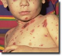
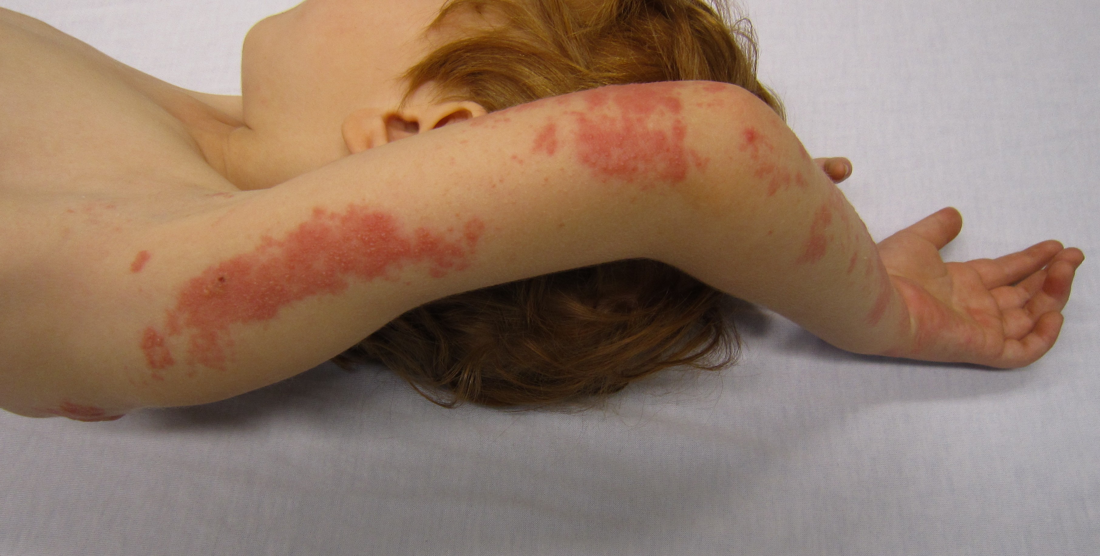
Image:Herpes zoster 6.jpg|Child with shingles who had a history of [[leukemia]] | |||
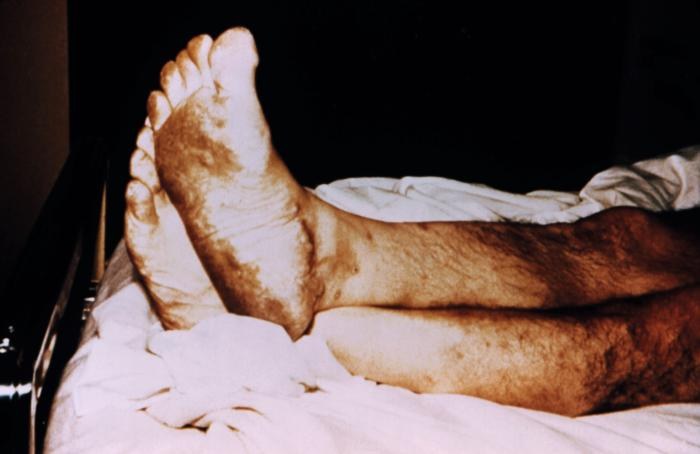
Image:Herpes zoster 7.jpg|[[Plantar]] foot [[rash]] caused by herpes zoster | |||
Image:Herpes zoster 8.jpg|[[Vesicle|Vesicular]] [[lesion]]s of Herpes zoster | |||
Image:Herpes zoster9.jpg|The [[pathological|pathologic]] changes seen on the surface of the right unilateral side of this elderly male patient’s tongue and chin, represent a herpes outbreak due to the [[Varicella zoster virus]] (VZV) pathogen. | |||
Image:Herpes zoster 10.jpg|The [[pustule|pustulo-]][[vesicle|vesicular]] [[rash]] on this African-American woman’s face, represents a herpes [[outbreak]] due to the [[Varicella zoster virus]] (VZV) pathogen. | |||
</gallery> | |||
<gallery> | |||
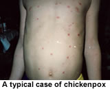
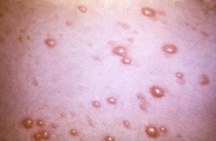
Image:Herpes zoster 11.jpg|This 1968 image depicted a number of varicella, or [[chickenpox]] [[lesion]]s on a patient’s back, which were displaying the characteristic “cropping” distribution, or manifesting themselves in clusters, each in a different developmental stage. | |||
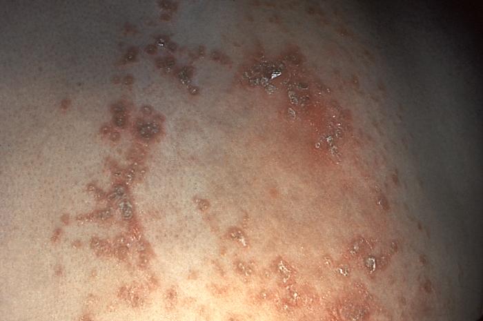
Image:Herpes zoster 12.jpg|This patient presented with what was differentially diagnosed as a herpes zoster [[outbreak]] in order to rule out [[syphilis]]. | |||
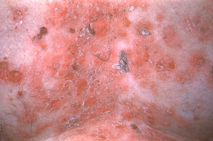
Image:Herpes zoster 13.jpg|This skin disorder was found to be herpes zoster, not [[Syphilis|syphilitic]] in nature as was initially suspected. | |||
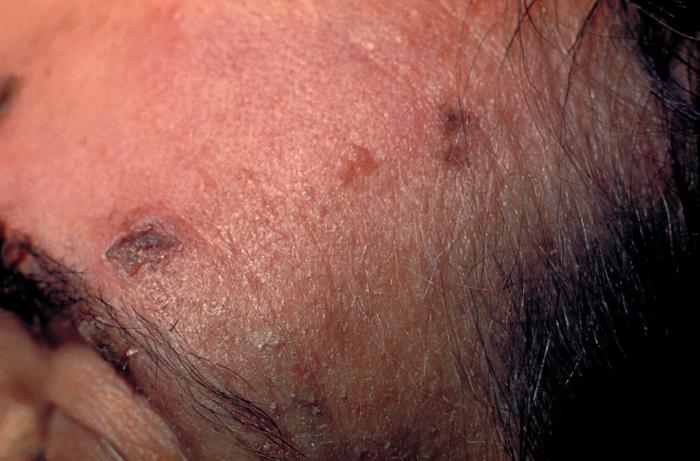
Image:Herpes zoster 14.jpg|These skin [[lesion]]s on the forehead of an elderly woman are due to the herpes zoster [[virus]] on the 21st day of the illness. | |||
Image:Herpes zoster 15.JPG|A case of shingles that demonstrates the typical [[dermatome|dermatomal]] distribution, in this case C8/T1 | |||
</gallery> | |||
====Head==== | =====Head===== | ||
<gallery> | <gallery> | ||
| Line 31: | Line 62: | ||
</gallery> | </gallery> | ||
====Trunk==== | =====Trunk===== | ||
<gallery> | <gallery> | ||
| Line 53: | Line 84: | ||
</gallery> | </gallery> | ||
====Extremities==== | =====Extremities===== | ||
<gallery> | <gallery> | ||
| Line 65: | Line 96: | ||
</gallery> | </gallery> | ||
====Neck==== | =====Neck===== | ||
<gallery> | <gallery> | ||
| Line 75: | Line 106: | ||
</gallery> | </gallery> | ||
====Genitourinary System==== | =====Genitourinary System===== | ||
<gallery> | <gallery> | ||
| Line 89: | Line 120: | ||
</gallery> | </gallery> | ||
====Skin==== | =====Skin===== | ||
(Images shown below courtesy of Charlie Goldberg, M.D., UCSD School of Medicine and VA Medical Center, San Diego, CA) | (Images shown below courtesy of Charlie Goldberg, M.D., UCSD School of Medicine and VA Medical Center, San Diego, CA) | ||
| Line 100: | Line 131: | ||
Image:Skin_zoster_buttocks3.jpg|Herpes Zoster: [[dermatome|Dermatomally]] distributed vesicles, many of which have coalesced, in patient with HZV infection. | Image:Skin_zoster_buttocks3.jpg|Herpes Zoster: [[dermatome|Dermatomally]] distributed vesicles, many of which have coalesced, in patient with HZV infection. | ||
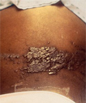
Image:Herpes zoster4.jpg|Shingles on waist | Image:Herpes zoster4.jpg|Shingles on waist | ||
</gallery> | |||
Image:Herpes zoster1.jpg|[[dermatome|Dermatomal]] involvement of [[rash]] | |||
Image:Herpes zoster2.jpg|[[dermatome|Dermatomal]] involvement of skin rash | |||
Image:Herpes zoster chest.jpg|Herpes zoster on the chest | |||
Image:Varicella child-ab.jpg | |||
Image:varicella zoster.jpg|Varicella zoster</gallery> | |||
</div> | </div> | ||
| Line 132: | Line 168: | ||
</gallery> | </gallery> | ||
</div> | </div> | ||
==References== | ==References== | ||
{{Reflist|2}} | {{Reflist|2}} | ||
Revision as of 12:19, 8 October 2014
|
Herpes zoster Microchapters |
|
Diagnosis |
|---|
|
History and Symptoms |
|
Treatment |
|
Case Studies |
|
Herpes zoster physical examination On the Web |
|
American Roentgen Ray Society Images of Herpes zoster physical examination |
|
Risk calculators and risk factors for Herpes zoster physical examination |
Editor-In-Chief: C. Michael Gibson, M.S., M.D. [1]; L. Katie Morrison, MD; Associate Editor(s)-In-Chief: Cafer Zorkun, M.D., Ph.D. [2]; Jesus Rosario Hernandez, M.D. [3].
Physical Examination
People with herpes zoster most commonly have a rash in one or two adjacent dermatomes (localized zoster). The rash most commonly appears on the trunk along a thoracic dermatome. The rash does not usually cross the body’s midline. However, approximately 20% of people have rash that overlaps adjacent dermatomes. Less commonly, the rash can be more widespread and affect three or more dermatomes. This condition is called disseminated zoster. This generally occurs only in people with compromised immune systems. Disseminated zoster can be difficult to distinguish from varicella.
The rash develops into clusters of clear vesicles. New vesicles continue to form over three to five days and progressively dry and crust over. They usually heal in two to four weeks. There may be permanent pigmentation changes and scarring on the skin.
Gallery
Skin
(Images shown below courtesy of Charlie Goldberg, M.D., UCSD School of Medicine and VA Medical Center, San Diego, CA)
-
-
Herpes Zoster: Dermatomally distributed vesicles, many of which have coalesced, in patient with HZV infection.
-
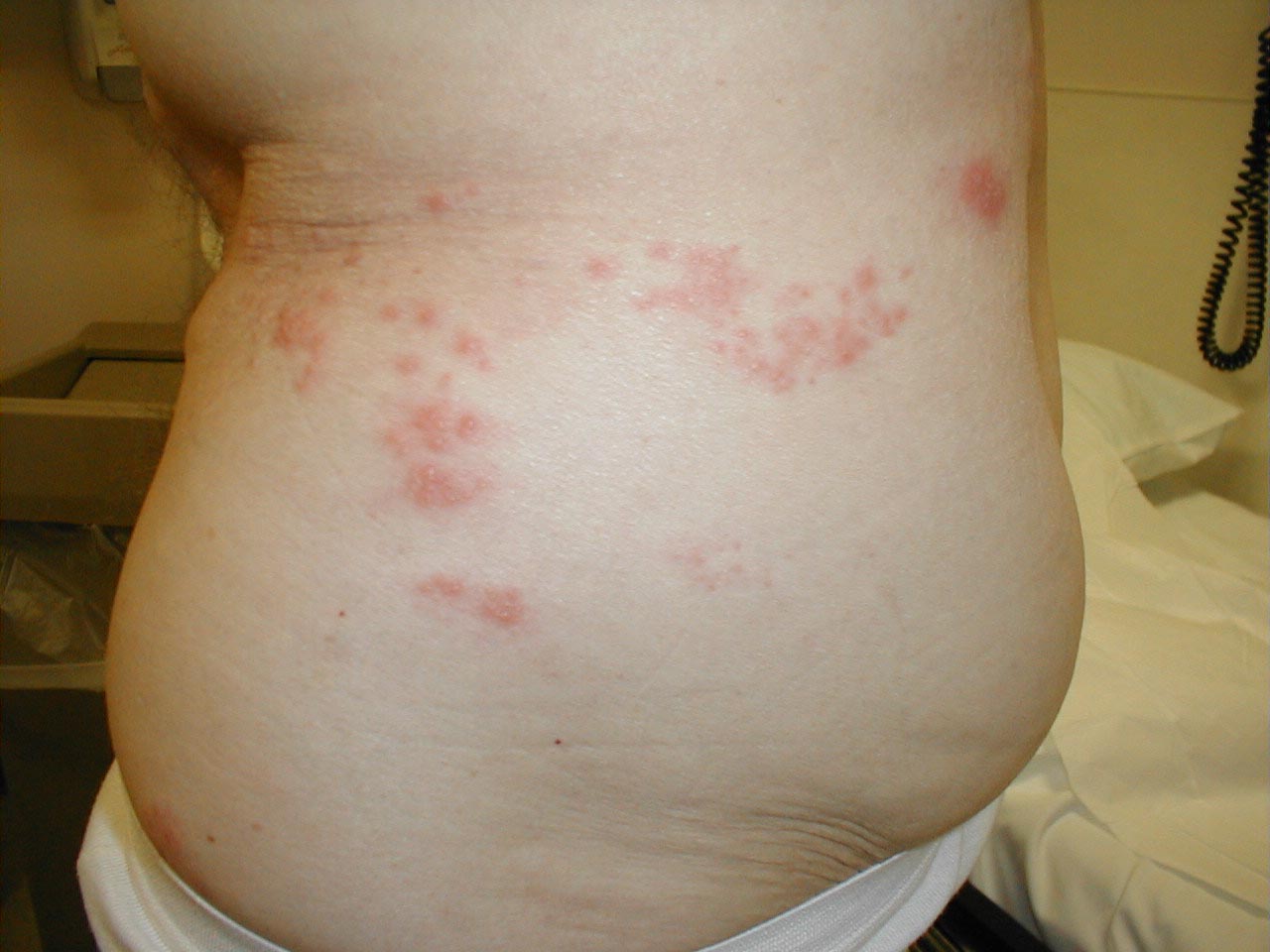
Herpes Zoster: Dermatomally distributed vesicles in patient with HZV infection.
-
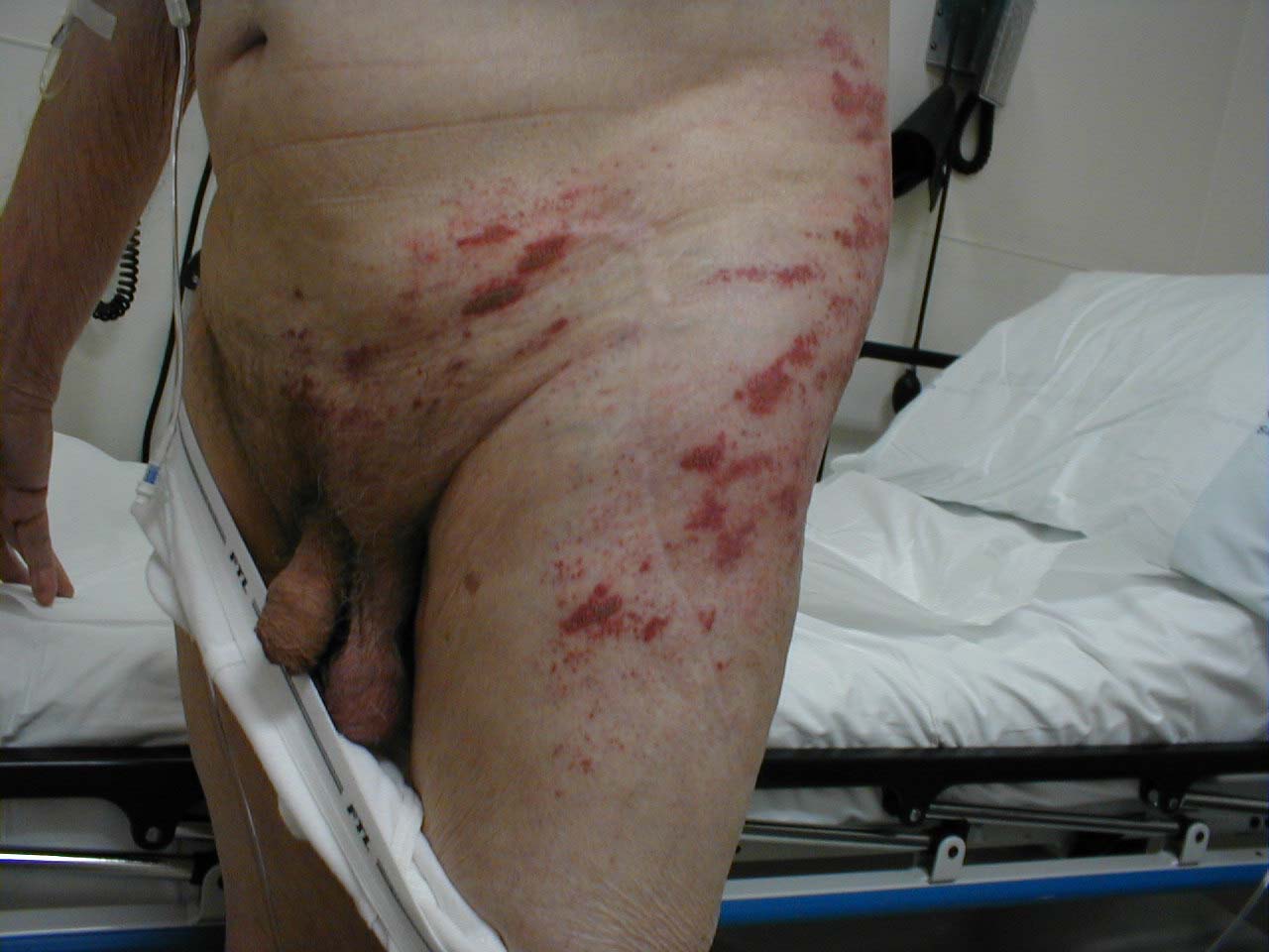
Herpes Zoster: Dermatomally distributed vesicles, many of which have coalesced, in patient with HZV infection.
-
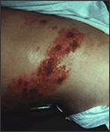
Shingles on waist
-
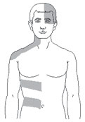
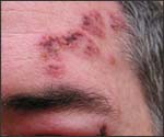
Dermatomal involvement of rash
-
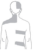
Dermatomal involvement of skin rash
-
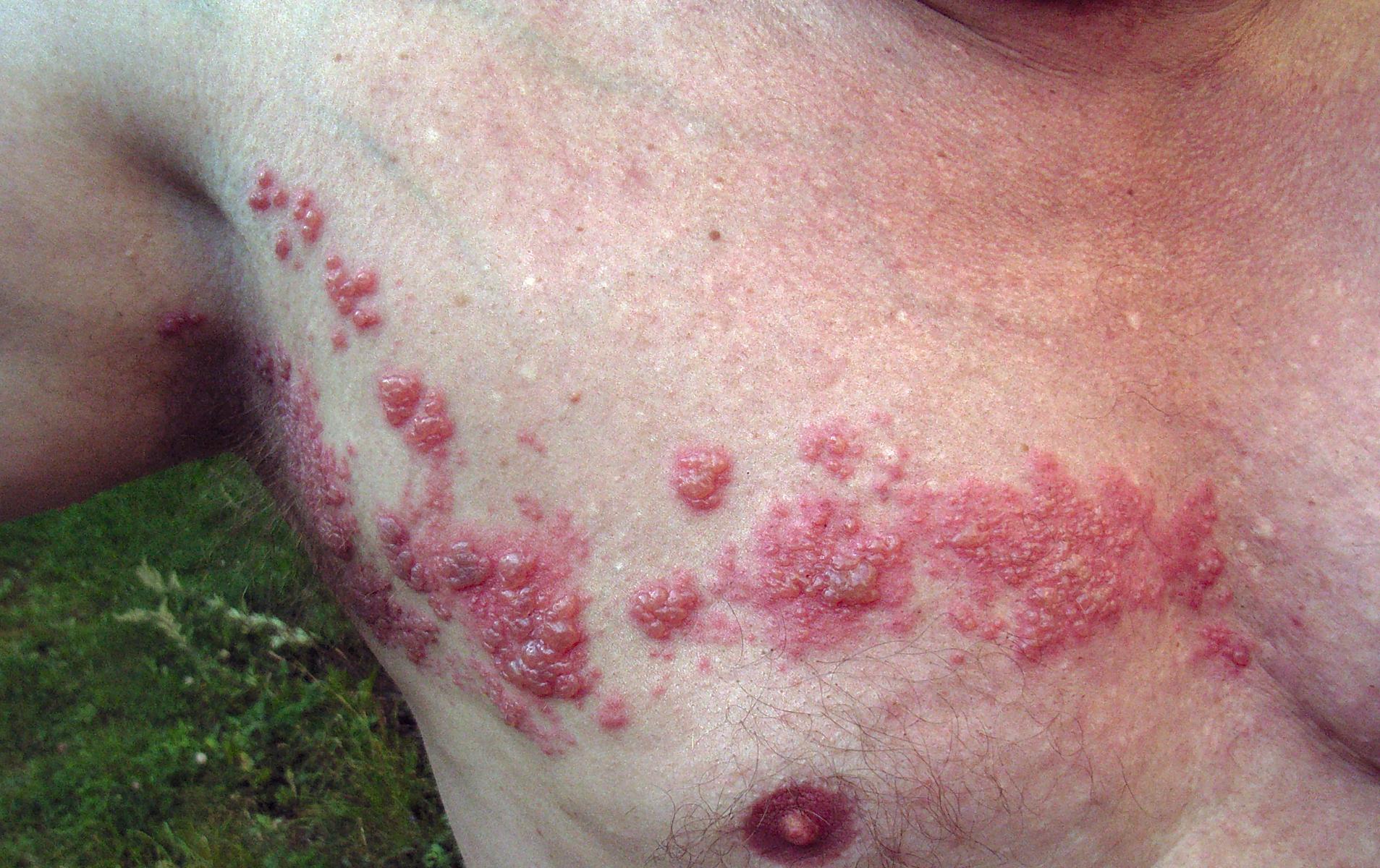
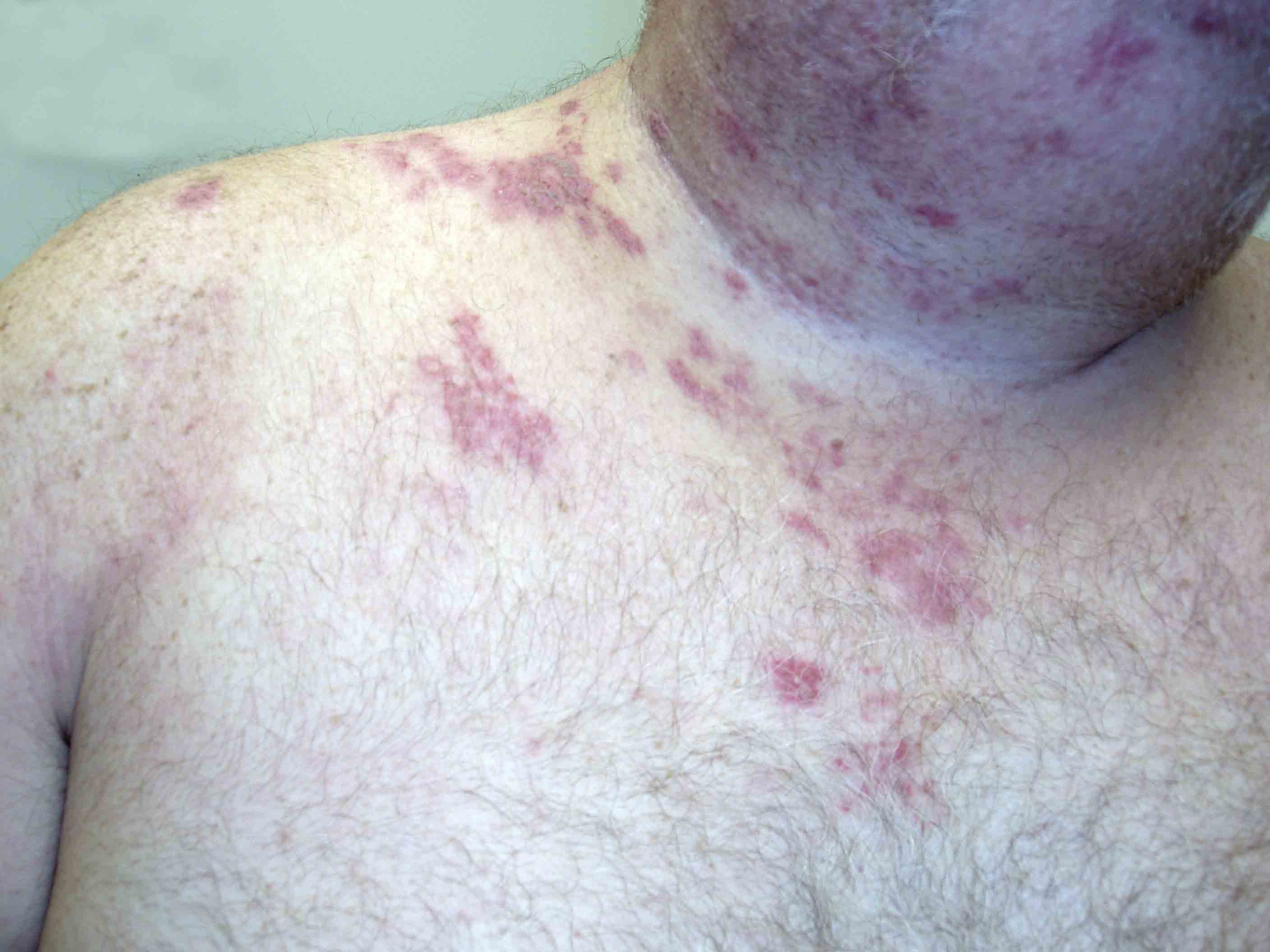
Herpes zoster on the chest
-
-
Varicella zoster


-
Herpes Zoster.
(Courtesy of Josh Fierer, M.D. and Charlie Goldberg, M.D.) -
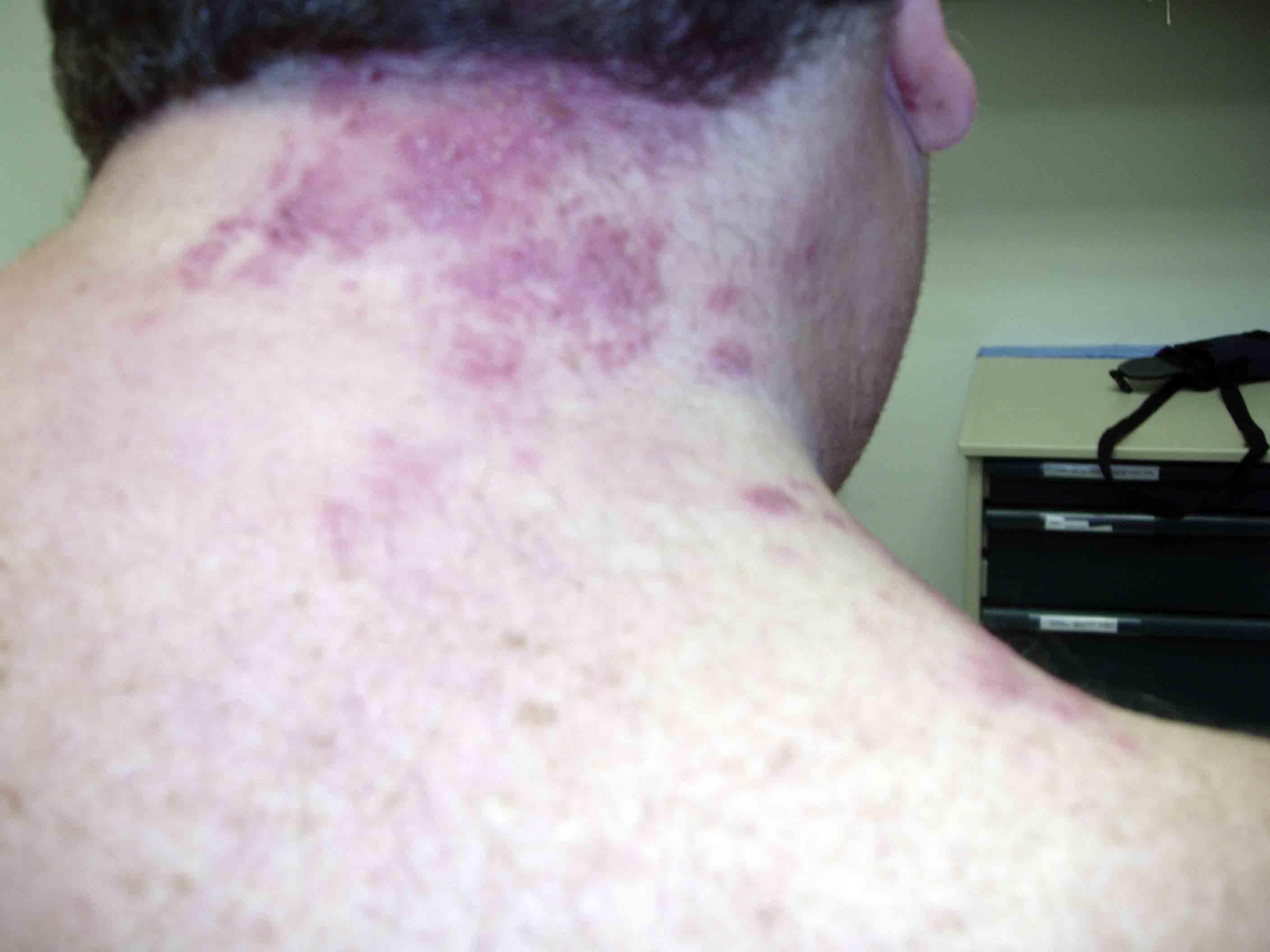
Herpes Zoster C3 Distribution: Dermatomally distributed vesicles, many of which have coalesced, in patient with HZV infection.
-
Herpes Zoster C3 Distribution: Dermatomally distributed vesicles, many of which have coalesced, in patient with HZV infection.
-
Shingles on face
-
Shingles on face


-
Child with shingles who had a history of leukemia
-
-
-
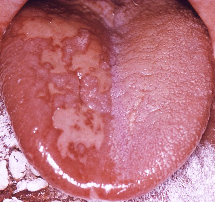
The pathologic changes seen on the surface of the right unilateral side of this elderly male patient’s tongue and chin, represent a herpes outbreak due to the Varicella zoster virus (VZV) pathogen.
-
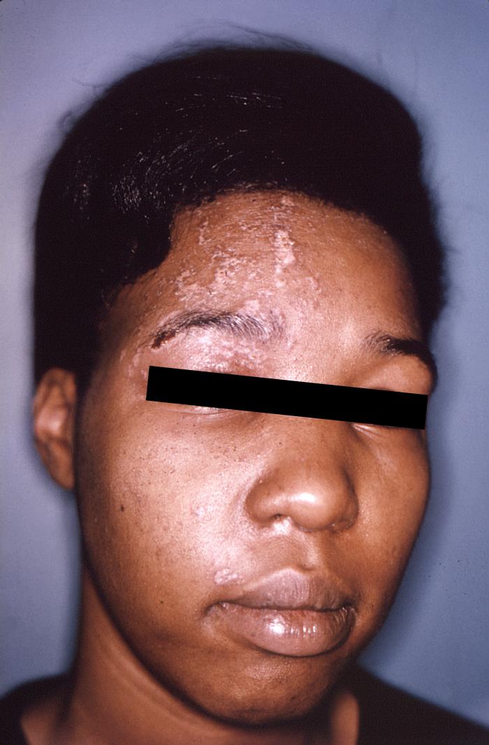
The pustulo-vesicular rash on this African-American woman’s face, represents a herpes outbreak due to the Varicella zoster virus (VZV) pathogen.


-
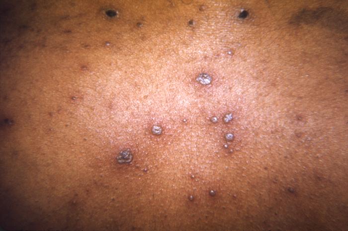
This 1968 image depicted a number of varicella, or chickenpox lesions on a patient’s back, which were displaying the characteristic “cropping” distribution, or manifesting themselves in clusters, each in a different developmental stage.
-
-
This skin disorder was found to be herpes zoster, not syphilitic in nature as was initially suspected.
-
-
A case of shingles that demonstrates the typical dermatomal distribution, in this case C8/T1


Head


Trunk


Extremities

Neck


Genitourinary System


Skin
(Images shown below courtesy of Charlie Goldberg, M.D., UCSD School of Medicine and VA Medical Center, San Diego, CA)
-
-
Herpes Zoster: Dermatomally distributed vesicles, many of which have coalesced, in patient with HZV infection.
-
Herpes Zoster: Dermatomally distributed vesicles in patient with HZV infection.
-
Herpes Zoster: Dermatomally distributed vesicles, many of which have coalesced, in patient with HZV infection.
-
Shingles on waist
-
Dermatomal involvement of rash
-
Dermatomal involvement of skin rash
-
Herpes zoster on the chest
-
-
Varicella zoster
-
Herpes Zoster.
(Courtesy of Josh Fierer, M.D. and Charlie Goldberg, M.D.) -
Herpes Zoster C3 Distribution: Dermatomally distributed vesicles, many of which have coalesced, in patient with HZV infection.
-
Herpes Zoster C3 Distribution: Dermatomally distributed vesicles, many of which have coalesced, in patient with HZV infection.
-
Shingles on face
-
Shingles on face
-
Child with shingles who had a history of leukemia
-
-
-
The pathologic changes seen on the surface of the right unilateral side of this elderly male patient’s tongue and chin, represent a herpes outbreak due to the Varicella zoster virus (VZV) pathogen.
-
The pustulo-vesicular rash on this African-American woman’s face, represents a herpes outbreak due to the Varicella zoster virus (VZV) pathogen.
-
This 1968 image depicted a number of varicella, or chickenpox lesions on a patient’s back, which were displaying the characteristic “cropping” distribution, or manifesting themselves in clusters, each in a different developmental stage.
-
-
This skin disorder was found to be herpes zoster, not syphilitic in nature as was initially suspected.
-
-
A case of shingles that demonstrates the typical dermatomal distribution, in this case C8/T1