First degree AV block pathophysiology: Difference between revisions
No edit summary |
|||
| Line 1: | Line 1: | ||
__NOTOC__ | __NOTOC__ | ||
{{First degree AV block}} | {{First degree AV block}} | ||
{{CMG}}; {{AE}} {{AEL}}, {{CZ}} | {{CMG}}; {{AE}} [[User:Mohammed Salih|Mohammed Salih, M.D.]], {{AEL}}, {{CZ}} | ||
==Overview== | ==Overview== | ||
Revision as of 21:53, 1 April 2020
|
First degree AV block Microchapters |
|
Diagnosis |
|---|
|
Treatment |
|
Case Studies |
|
First degree AV block pathophysiology On the Web |
|
American Roentgen Ray Society Images of First degree AV block pathophysiology |
|
Risk calculators and risk factors for First degree AV block pathophysiology |
Editor-In-Chief: C. Michael Gibson, M.S., M.D. [1]; Associate Editor(s)-in-Chief: Mohammed Salih, M.D., Ahmed Elsaiey, MBBCH [2], Cafer Zorkun, M.D., Ph.D. [3]
Overview
The atrioventricular node is a normal electrical pathway between the atria and ventricles and it is located in the right atrium. First degree AV block pathogenesis can be attributed to an electrical conduction delay in the AV node or His-Purkinje system. First degree AV block can be associated with normal QRS complex or wide QRS complex on the ECG.
Pathophysiology
Physiology
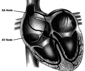
- The atrioventricluar node is the normal electrical pathway between the atria and ventricles. It is located in the right atrium near to the tricuspid valve leaflet.
- The AV node receives blood supply from the right coronary artery in most of the population.
- The bundle of His is the electrical connection from the AV node to the interventricular septum. At the end of the septum, bundle of His is divided into the right and left bundle branches to the ventricular walls.
Pathogenesis
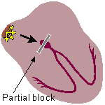
- First degree AV block broadly means the prolongation of the PR interval on the ECG with normal atrioventricular node function.
- First degree AV block pathogenesis can be attributed to an electrical conduction delay in one of the following:[1]
- Atrioventricular node
- His-Purkinje system which is formed of both bundle of His and Purkinje fibers
- The conduction delay can be also in both of the AV node and His-Purkinje system.
- One of the characteristics of first degree AV block that there are no beats skipped and it has a regular rhythm.
First Degree AV Block with Normal QRS Duration
First degree AV block with normal QRS duration, in majority of cases, results from atrial or AV nodal delay. Other probable sites include the bundle of His and infra-Hisian conduction system.
First Degree AV Block with Wide QRS Complex
First degree AV block with wide QRS complex most often results from delay in conduction in the bundle of His and in some patients, the AV node.
References
- ↑ Lewalter T, Pürerfellner H, Ungar A, Rieger G, Mangoni L, Duru F; et al. (2018). ""First-degree AV block-a benign entity?" Insertable cardiac monitor in patients with 1st-degree AV block reveals presence or progression to higher grade block or bradycardia requiring pacemaker implant". J Interv Card Electrophysiol. 52 (3): 303–306. doi:10.1007/s10840-018-0439-7. PMID 30105427.

