Fanconi anemia physical examination
|
Fanconi anemia Microchapters |
|
Diagnosis |
|---|
|
Treatment |
|
Case Studies |
|
Fanconi anemia physical examination On the Web |
|
American Roentgen Ray Society Images of Fanconi anemia physical examination |
|
Risk calculators and risk factors for Fanconi anemia physical examination |
Editor-In-Chief: C. Michael Gibson, M.S., M.D. [1]; Associate Editor(s)-in-Chief:
Overview
CLINICAL FEATURES
Congenital anomalies — Congenital malformations are the most common presenting features of FA. Malformations are reported in 60 to 75 percent of patients, but many in the field believe this represents an underestimate, as many patients with FA do not manifest classical physical findings.
Young adults with more subtle clinical findings increasingly may be identified from genomic sequencing. Despite the high frequency of malformations, only a small percentage of patients with FA (<5 percent) are diagnosed within the first year of life based on classic congenital anomalies. Thus, while the presence of these findings provides an important clue to the diagnosis, their absence does not eliminate the possibility of FA.
In a series of 370 patients enrolled in the International FA Registry and a review of over 2000 patients reported in the literature from 1927 to 2009, the most common developmental abnormalities included the following [20,68]:
● Skin findings (approximately 40 to 60 percent), including hyper- or hypopigmentation or café-au-lait spots
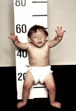
● Short stature (40 to 60 percent)
● Thumb or other radial ray abnormalities (50 percent)
• Thumbs absent or hypoplastic, bifid/duplicated, rudimentary, triphalangeal (35 percent)
• Radii absent or hypoplastic (7 percent)
• Hands/other such as flat thenar eminence, clinodactyly, polydactyly, missing first metacarpal, dysplastic ulnae (6 percent)
● Axial skeletal abnormalities (25 percent), especially microcephaly, triangular facies, short/webbed neck, vertebral anomalies
● Eye malformations (20 to 40 percent), including strabismus and hypo/hypertelorism
● Renal and urinary tract malformations (approximately 20 to 30 percent) including horseshoe, ectopic, dysplastic, or absent kidney; hydronephrosis; hydroureter
● Gonadal/Genital malformations
• In males, hypospadias, micropenis, undescended/absent testes, infertility (25 percent)
• In females, uterus malformation, small ovaries, hypogenitalia (<5 percent)
● Ear abnormalities (10 to 20 percent) with conductive hearing loss due to middle ear anomalies or atretic ear canal
● Congenital heart disease (approximately 5 percent) such as patent ductus arteriosus, ventricular septal defect, aortic coarctation, truncus arteriosus
● Gastrointestinal anomalies (approximately 5 percent) such as tracheoesophageal fistula, esophageal atresia, intestinal atresia, imperforate anus
● Central nervous system abnormalities (<5 percent) involving the pituitary gland (eg, small, interrupted pituitary stalk syndrome), hydrocephalus, cerebellar hypoplasia, or absent corpus callosumPatients with [disease name] usually appear [general appearance]. Physical examination of patients with [disease name] is usually remarkable for [finding 1], [finding 2], and [finding 3].
OR
Common physical examination findings of [disease name] include [finding 1], [finding 2], and [finding 3].
OR
The presence of [finding(s)] on physical examination is diagnostic of [disease name].
OR
The presence of [finding(s)] on physical examination is highly suggestive of [disease name].
Physical Examination
- Physical examination of patients with [disease name] is usually normal.
OR
- Physical examination of patients with [disease name] is usually remarkable for:[finding 1], [finding 2], and [finding 3].
- The presence of [finding(s)] on physical examination is diagnostic of [disease name].
- The presence of [finding(s)] on physical examination is highly suggestive of [disease name].
Appearance of the Patient
- Patients with [disease name] usually appear [general appearance].
Vital Signs
- High-grade / low-grade fever
- Hypothermia / hyperthermia may be present
- Tachycardia with regular pulse or (ir)regularly irregular pulse
- Bradycardia with regular pulse or (ir)regularly irregular pulse
- Tachypnea / bradypnea
- Kussmal respirations may be present in _____ (advanced disease state)
- Weak/bounding pulse / pulsus alternans / paradoxical pulse / asymmetric pulse
- High/low blood pressure with normal pulse pressure / wide pulse pressure / narrow pulse pressure
Skin
- Skin examination of patients with [disease name] is usually normal.
OR
-
Description (Adapted from Dermatology Atlas)
-
Description (Adapted from Dermatology Atlas)

HEENT
- HEENT examination of patients with [disease name] is usually normal.
OR
- Abnormalities of the head/hair may include ___
- Evidence of trauma
- Icteric sclera
- Nystagmus
- Extra-ocular movements may be abnormal
- Pupils non-reactive to light / non-reactive to accommodation / non-reactive to neither light nor accommodation
- Ophthalmoscopic exam may be abnormal with findings of ___
- Hearing acuity may be reduced
- Weber test may be abnormal (Note: A positive Weber test is considered a normal finding / A negative Weber test is considered an abnormal finding. To avoid confusion, you may write "abnormal Weber test".)
- Rinne test may be positive (Note: A positive Rinne test is considered a normal finding / A negative Rinne test is considered an abnormal finding. To avoid confusion, you may write "abnormal Rinne test".)
- Exudate from the ear canal
- Tenderness upon palpation of the ear pinnae/tragus (anterior to ear canal)
- Inflamed nares / congested nares
- Purulent exudate from the nares
- Facial tenderness
- Erythematous throat with/without tonsillar swelling, exudates, and/or petechiae
Neck
- Neck examination of patients with [disease name] is usually normal.
OR
- Jugular venous distension
- Carotid bruits may be auscultated unilaterally/bilaterally using the bell/diaphragm of the otoscope
- Lymphadenopathy (describe location, size, tenderness, mobility, and symmetry)
- Thyromegaly / thyroid nodules
- Hepatojugular reflux
Lungs
- Pulmonary examination of patients with [disease name] is usually normal.
OR
- Asymmetric chest expansion / Decreased chest expansion
- Lungs are hypo/hyperresonant
- Fine/coarse crackles upon auscultation of the lung bases/apices unilaterally/bilaterally
- Rhonchi
- Vesicular breath sounds / Distant breath sounds
- Expiratory/inspiratory wheezing with normal / delayed expiratory phase
- Wheezing may be present
- Egophony present/absent
- Bronchophony present/absent
- Normal/reduced tactile fremitus
Heart
- Cardiovascular examination of patients with [disease name] is usually normal.
OR
- Chest tenderness upon palpation
- PMI within 2 cm of the sternum (PMI) / Displaced point of maximal impulse (PMI) suggestive of ____
- Heave / thrill
- Friction rub
- S1
- S2
- S3
- S4
- Gallops
- A high/low grade early/late systolic murmur / diastolic murmur best heard at the base/apex/(specific valve region) may be heard using the bell/diaphgram of the otoscope
Abdomen
Abdominal examination of patients with [disease name] is usually normal.
OR
- Abdominal distention
- Abdominal tenderness in the right/left upper/lower abdominal quadrant
- Rebound tenderness (positive Blumberg sign)
- A palpable abdominal mass in the right/left upper/lower abdominal quadrant
- Guarding may be present
- Hepatomegaly / splenomegaly / hepatosplenomegaly
- Additional findings, such as obturator test, psoas test, McBurney point test, Murphy test
Back
- Back examination of patients with [disease name] is usually normal.
OR
- Point tenderness over __ vertebrae (e.g. L3-L4)
- Sacral edema
- Costovertebral angle tenderness bilaterally/unilaterally
- Buffalo hump
Genitourinary
- Genitourinary examination of patients with [disease name] is usually normal.
OR
- A pelvic/adnexal mass may be palpated
- Inflamed mucosa
- Clear/(color), foul-smelling/odorless penile/vaginal discharge
Neuromuscular
- Neuromuscular examination of patients with [disease name] is usually normal.
OR
- Patient is usually oriented to persons, place, and time
- Altered mental status
- Glasgow coma scale is ___ / 15
- Clonus may be present
- Hyperreflexia / hyporeflexia / areflexia
- Positive (abnormal) Babinski / plantar reflex unilaterally/bilaterally
- Muscle rigidity
- Proximal/distal muscle weakness unilaterally/bilaterally
- ____ (finding) suggestive of cranial nerve ___ (roman numerical) deficit (e.g. Dilated pupils suggestive of CN III deficit)
- Unilateral/bilateral upper/lower extremity weakness
- Unilateral/bilateral sensory loss in the upper/lower extremity
- Positive straight leg raise test
- Abnormal gait (describe gait: e.g. ataxic (cerebellar) gait / steppage gait / waddling gait / choeiform gait / Parkinsonian gait / sensory gait)
- Positive/negative Trendelenburg sign
- Unilateral/bilateral tremor (describe tremor, e.g. at rest, pill-rolling)
- Normal finger-to-nose test / Dysmetria
- Absent/present dysdiadochokinesia (palm tapping test)
Extremities
- Extremities examination of patients with [disease name] is usually normal.
OR
- Clubbing
- Cyanosis
- Pitting/non-pitting edema of the upper/lower extremities
- Muscle atrophy
- Fasciculations in the upper/lower extremity