Ependymoma pathophysiology: Difference between revisions
| Line 33: | Line 33: | ||
===Associated Conditions=== | ===Associated Conditions=== | ||
The subependymal giant-cell astrocytoma, also called giant-cell glioma, is typically associated with [[tuberous sclerosis]] but can occur | The subependymal giant-cell astrocytoma, also called giant-cell glioma, is typically associated with [[tuberous sclerosis]] but can occur independently. | ||
==References== | ==References== | ||
Revision as of 16:11, 6 October 2015
|
Ependymoma Microchapters |
|
Diagnosis |
|---|
|
Treatment |
|
Case Studies |
|
Ependymoma pathophysiology On the Web |
|
American Roentgen Ray Society Images of Ependymoma pathophysiology |
|
Risk calculators and risk factors for Ependymoma pathophysiology |
Editor-In-Chief: C. Michael Gibson, M.S., M.D. [1] Associate Editor(s)-in-Chief: Ahmad Al Maradni, M.D. [2]
Overview
Pathology
- Ependymomas represent a relatively broad group of glial tumours which share common origin from differentiated ependymal cells lining the ventricles of the brain or the central canal of the spinal cord. They account for ~5% of all neuroepithelial neoplasms, ~10% of all paediatric brain tumours and up to 33% of brain tumours occurring in those less than 3 years of age.
- Ependymomas can occur anywhere, but certain location are typical. Common locations include:
- Floor of the fourth ventricle (common location in children)
- Spinal cord ependymoma
- Myxopapillary ependymoma (conus medullaris)
- Supratentorial ependymoma
- The subependymomas, variant of the ependymoma, arise in the fourth ventricle but may occur in the septum pellucidum and the cervical spinal cord.
Gross Pathology
- Ependymomas are well-encapsulated tumors and usually arise from the floor of the fourth ventricle, situated in the lower back portion of the brain.
-
![Fourth ventricle ependymomas frequently extend out of the ventricle into the subarachnoid space.[1]](/images/0/0f/Epyndemomas_gross.jpg)
Fourth ventricle ependymomas frequently extend out of the ventricle into the subarachnoid space.[1]
![Fourth ventricle ependymomas frequently extend out of the ventricle into the subarachnoid space.[1]](/index.php/File:Epyndemomas_gross.jpg)
Microscopic Pathology
- Ependymomas are composed of cells with regular, round to oval nuclei. There is a variably dense fibrillary background.
- Tumor cells may form gland-like round or elongated structures that resemble the embryologic ependymal canal, with long, delicate processes extending into the lumen; more frequently present are perivascular pseudorosettes in which tumor cells are arranged around vessels with an intervening zone consisting of thin ependymal processes directed toward the wall of the vessel.[2].
- Ependymal rosettes are rare but pathognomonic feature.
-
![Micrograph of an ependymoma. H&E stain show pseudoressettes pattern of ependymal cells.[3]](/images/0/0a/532px-Ependymoma_low_intermed_mag.jpg)
Micrograph of an ependymoma. H&E stain show pseudoressettes pattern of ependymal cells.[3]
-
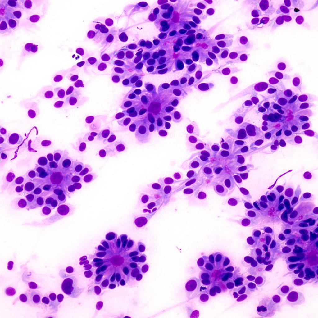
True ependymal rosette consisting of tumor cells arranged around well-defined lumens forming gland-like structures.<ref name=radiopaedia> Image courtesy of Dr Dharam RamnaniRadiopaedia (original file
-
![Higher magnification of one of the true ependymal rosettes showing a collar of cells around a central lumen forming a gland-like structure.[4]](/images/b/b6/Ependymoma-true-ependymal-rosettes123.jpg)
Higher magnification of one of the true ependymal rosettes showing a collar of cells around a central lumen forming a gland-like structure.[4]
![Micrograph of an ependymoma. H&E stain show pseudoressettes pattern of ependymal cells.[3]](/index.php/File:532px-Ependymoma_low_intermed_mag.jpg)

![Higher magnification of one of the true ependymal rosettes showing a collar of cells around a central lumen forming a gland-like structure.[4]](/index.php/File:Ependymoma-true-ependymal-rosettes123.jpg)
Associated Conditions
The subependymal giant-cell astrocytoma, also called giant-cell glioma, is typically associated with tuberous sclerosis but can occur independently.
References
- ↑ EPENDYMOMA. http://librepathology.org/wiki/index.php/File:AFIP405713G-EPENDYMOMA.jpg 2015. URL Accessed on 10 6, 2015
- ↑ Kumar, et al. (2005). The Central Nervous System. Pathologic Basis of Disease. 7th Edition. Philadelphia: Elsevier Saunders.
- ↑ Ependymoma. https://en.wikipedia.org/wiki/Ependymoma#/media/File:Ependymoma_low_intermed_mag.jpg. URL Accessed on 10 6, 2015
- ↑ Image courtesy of Dr Dharam RamnaniRadiopaedia (original file‘’here’’).[http://radiopaedia.org/licence CreativeCommons BY-SANC