Diverticulitis x ray: Difference between revisions
Jump to navigation
Jump to search
| Line 18: | Line 18: | ||
**Multiple air and fluid levels in case perforation occurs. | **Multiple air and fluid levels in case perforation occurs. | ||
[[Image:866011f1abdb744127ddd20d04acff jumbo.jpeg|Center|500px]] | [[Image:866011f1abdb744127ddd20d04acff jumbo.jpeg|Center|500px]] | ||
[[Image:Diverticulitis abdominal x ray.jpeg|Center|500px]] | |||
*The single contrast technique may be preferred over the double contrast technique in the following cases: | *The single contrast technique may be preferred over the double contrast technique in the following cases: | ||
**The patient is unable to turn quickly/effectively | **The patient is unable to turn quickly/effectively | ||
Revision as of 20:32, 9 June 2017
|
Diverticulitis Microchapters |
|
Diagnosis |
|---|
|
Treatment |
|
Case Studies |
|
Diverticulitis x ray On the Web |
|
American Roentgen Ray Society Images of Diverticulitis x ray |
Editor-In-Chief: C. Michael Gibson, M.S., M.D. [1] Associate Editor(s)-in-Chief:
Overview
X Ray
Abdominal X ray
Barium enema
- X-ray barium enema is not the first diagnostic procedure to diagnose acute diverticulitis. However, it may be useful in case the CT scan is not available.[1][2][3][4]
- Barium enema was being used in diagnosis of acute diverticulitis but it was not the best procedure to diagnose the disease. Enema has many disadvantages which include:[5]
- Enema rupture
- This rupture may cause cellulitis and peritonitis.
- If failed, it will lead to the delay of the other imaging procedures like CT scan, endoscopy and angiography to be held.
- It may cause acute intestinal obstruction.
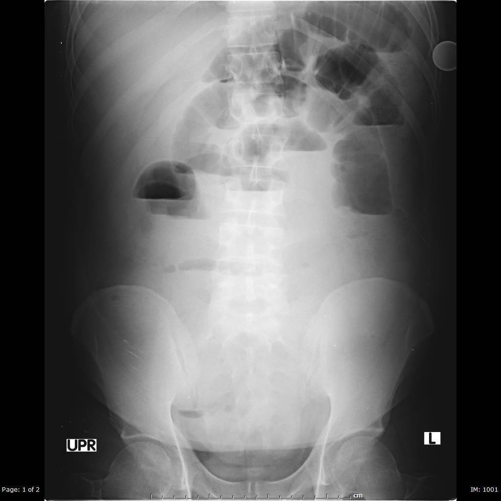
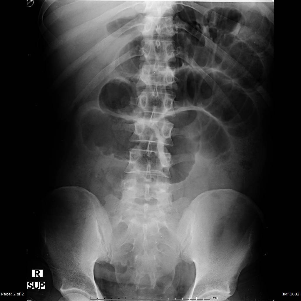
- The radiological findings in the abdominal x ray includes the following:
- It shows intestinal obstruction
- Multiple air and fluid levels in case perforation occurs.
- The single contrast technique may be preferred over the double contrast technique in the following cases:
- The patient is unable to turn quickly/effectively
- Double contrast technique requires rapid changes in patient position
- When only the position and length of a stricture is required
- Evaluation for acute diverticulitis when the CT is unavailable for whatever reason)
- Evaluating for a colonic fistula
- Evaluation for postoperative leak after colon surgery
- Contraindications of the barium enema include the following:[6]
- Patients with pnemoperitoneum shown in the chest X ray.
- Patients who did a recent deep rectal biopsy.
Chest X ray
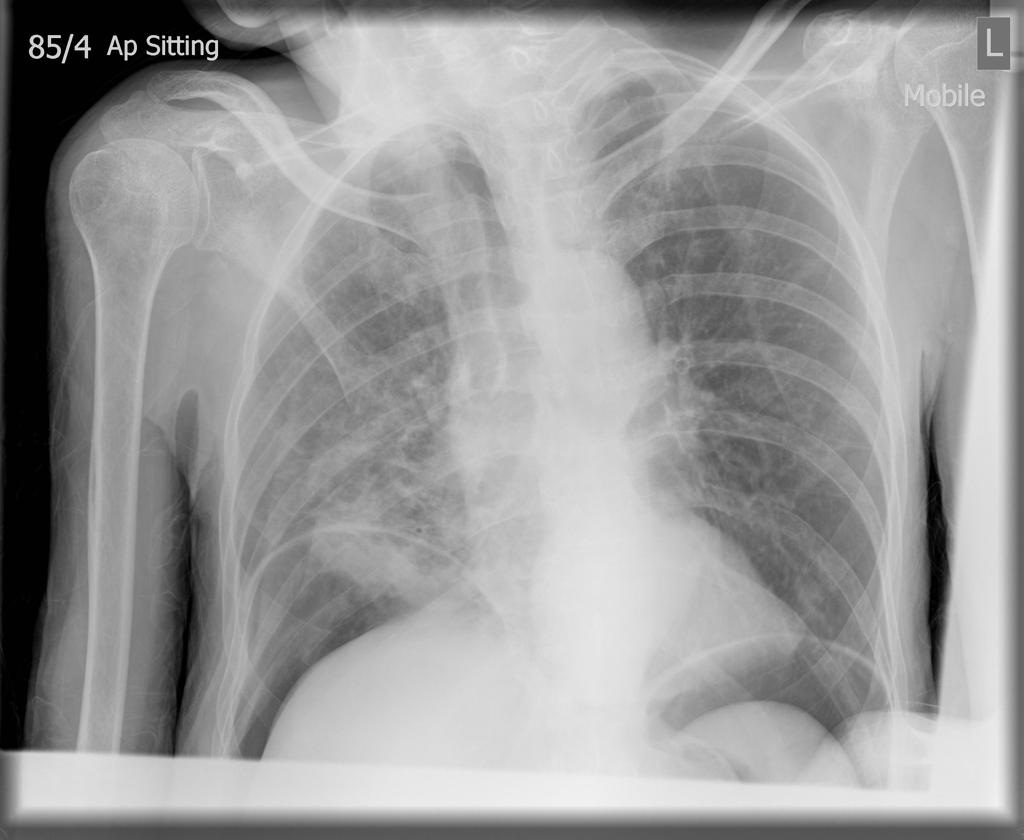
- In small percentage (about 27-33%) of the acute abdomen patients, including diverticulitis, they may have a respiratory abnormality. Hence, chest x ray is recommended in patients suspected with diverticulitis.
- Chest X ray in cases of diverticulitis could show pneumoperitoneum which is a gas within the peritoneal cavity, and is often the harbinger of a critical illness. Discovering this abnormality will lead to change in the case manangement and the chest X ray is the best radio-modality which can show the pneumoperitoneum.[7]
References
- ↑ Rafferty J, Shellito P, Hyman NH, Buie WD, Standards Committee of American Society of Colon and Rectal Surgeons (2006). "Practice parameters for sigmoid diverticulitis". Dis Colon Rectum. 49 (7): 939–44. doi:10.1007/s10350-006-0578-2. PMID 16741596.
- ↑ Doris PE, Strauss RW (1985). "The expanded role of the barium enema in the evaluation of patients presenting with acute abdominal pain". J Emerg Med. 3 (2): 93–100. PMID 4093571.
- ↑ McKee RF, Deignan RW, Krukowski ZH (1993). "Radiological investigation in acute diverticulitis". Br J Surg. 80 (5): 560–5. PMID 8518890.
- ↑ Hayward MW, Hayward C, Ennis WP, Roberts CJ (1984). "A pilot evaluation of radiography of the acute abdomen". Clin Radiol. 35 (4): 289–91. PMID 6734062.
- ↑ Gottesman L, Zevon SJ, Brabbee GW, Dailey T, Wichern WA (1984). "The use of water-soluble contrast enemas in the diagnosis of acute lower left quadrant peritonitis". Dis Colon Rectum. 27 (2): 84–8. PMID 6697835.
- ↑ Harned RK, Consigny PM, Cooper NB, Williams SM, Woltjen AJ (1982). "Barium enema examination following biopsy of the rectum or colon". Radiology. 145 (1): 11–6. doi:10.1148/radiology.145.1.7122864. PMID 7122864.
- ↑ Field S, Guy PJ, Upsdell SM, Scourfield AE (1985). "The erect abdominal radiograph in the acute abdomen: should its routine use be abandoned?". Br Med J (Clin Res Ed). 290 (6486): 1934–6. PMC 1416036. PMID 3924315.


