Craniopharyngioma classification
|
Craniopharyngioma Microchapters |
|
Diagnosis |
|---|
|
Treatment |
|
Case Studies |
|
Craniopharyngioma classification On the Web |
|
American Roentgen Ray Society Images of Craniopharyngioma classification |
|
Risk calculators and risk factors for Craniopharyngioma classification |
Editor-In-Chief: C. Michael Gibson, M.S., M.D. [1]Associate Editor(s)-in-Chief: Marjan Khan M.B.B.S.[2]
Overview
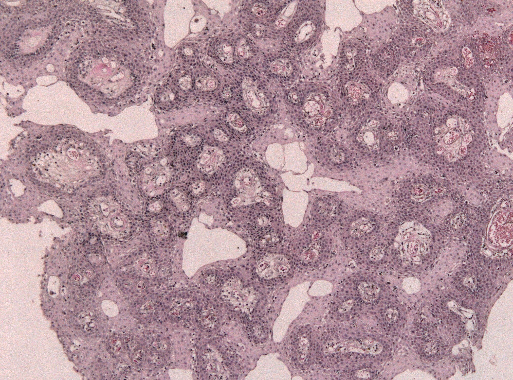
There are two subtypes of craniopharyngioma based on the histological and imaging features: adamantinomatous and papillary.
Classification
- Craniopharyngiomas are believed to be derive from Rathke cleft rather than squamous cell rests. [1]
- Craniopharyngiomas are classified according to their histologic appearance.
- The histological appearances of the two pathological subtypes are different, accounting for the different imaging features.
- The subtypes are said to differ not only in appearances, but also in prognosis and epidemiology.[2]
- Adamantinomatous
 |
- This type is seen predominantly in children.[1]
- It consists of reticular epithelial cells which have appearances reminiscent of the enamel pulp of developing teeth.
- There may be single or multiple cysts filled with thick oily fluid high in protein, blood products, and/or cholesterol, creating the so called "machinery oil".[3]
- "Wet keratin nodules" are a characteristic histological feature.[4]
- Calcification is usually present in 90% of the cases.
- They are more locally aggressive.
- It has higher rate of recurrence.
- Activating beta-catenin gene mutations are found in adamantinomatous tumors.[5]
- Papillary
 |
- The papillary subtype is seen almost exclusively in adults.[1]
- It is formed of masses of metaplastic squamous cells.[4]
- Mixed
- 15% of them share imaging features and prognosis similar to the adamantinomatous subtye.[7]
References
- ↑ 1.0 1.1 1.2 Mortini P (August 2017). "Craniopharyngiomas: a life-changing tumor". Endocrine. 57 (2): 191–192. doi:10.1007/s12020-016-1192-2. PMID 27981519.
- ↑ Classification of Craniopharyngioma. Cancer gov. http://www.cancer.gov/types/brain/hp/child-cranio-treatment-pdq#link/_40_toc
- ↑ 3.0 3.1 Müller HL (April 2017). "Risk-adapted, long-term management in childhood-onset craniopharyngioma". Pituitary. 20 (2): 267–281. doi:10.1007/s11102-016-0751-0. PMID 27604996.
- ↑ 4.0 4.1 4.2 Petito CK, DeGirolami U, Earle KM (April 1976). "Craniopharyngiomas: a clinical and pathological review". Cancer. 37 (4): 1944–52. PMID 1260697.
- ↑ Duff J, Meyer FB, Ilstrup DM, Laws ER, Schleck CD, Scheithauer BW (February 2000). "Long-term outcomes for surgically resected craniopharyngiomas". Neurosurgery. 46 (2): 291–302, discussion 302–5. PMID 10690718.
- ↑ Bunin GR, Surawicz TS, Witman PA, Preston-Martin S, Davis F, Bruner JM (October 1998). "The descriptive epidemiology of craniopharyngioma". J. Neurosurg. 89 (4): 547–51. doi:10.3171/jns.1998.89.4.0547. PMID 9761047.
- ↑ Sekine S, Takata T, Shibata T; et al. (2004). "Expression of enamel proteins and LEF1 in adamantinomatous craniopharyngioma: evidence for its odontogenic epithelial differentiation". Histopathology. 45 (6): 573–9. doi:10.1111/j.1365-2559.2004.02029.x. PMID 15569047. Unknown parameter
|month=ignored (help)