Craniopharyngioma MRI: Difference between revisions
Jump to navigation
Jump to search
(→MRI) |
|||
| Line 7: | Line 7: | ||
===Adamantinomatous=== | ===Adamantinomatous=== | ||
The findings on | The findings on MRI are: | ||
*'''Cysts''' | *'''Cysts''' | ||
**Variable but 80% are mostly or partly T2 hyperintense | **Variable but 80% are mostly or partly T2 hyperintense | ||
| Line 21: | Line 21: | ||
'''MR spectroscopy''': Cyst contents may show a broad lipid spectrum, with an otherwise flat baseline. | '''MR spectroscopy''': Cyst contents may show a broad lipid spectrum, with an otherwise flat baseline. | ||
===Papillary=== | |||
The findings on MRI are: | |||
*'''Cysts''' | |||
**When present they are variable in signal | |||
**85% T1 hypointense | |||
*Solid component | |||
**T1: iso- to lightly hypointense to brain | |||
**T1 C+: vivid enhancement | |||
**T2: variable/mixed | |||
'''MR spectroscopy''': cyst contents does not show a broad lipid spectrum as they are filled with water fluid | |||
([http://www.radswiki.net Images courtesy of RadsWiki]) | ([http://www.radswiki.net Images courtesy of RadsWiki]) | ||
Revision as of 00:56, 23 August 2015
|
Craniopharyngioma Microchapters |
|
Diagnosis |
|---|
|
Treatment |
|
Case Studies |
|
Craniopharyngioma MRI On the Web |
|
American Roentgen Ray Society Images of Craniopharyngioma MRI |
Editor-In-Chief: C. Michael Gibson, M.S., M.D. [1]
Overview
MRI
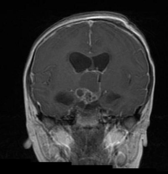
Adamantinomatous
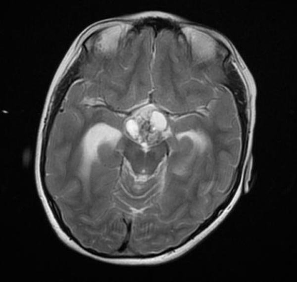
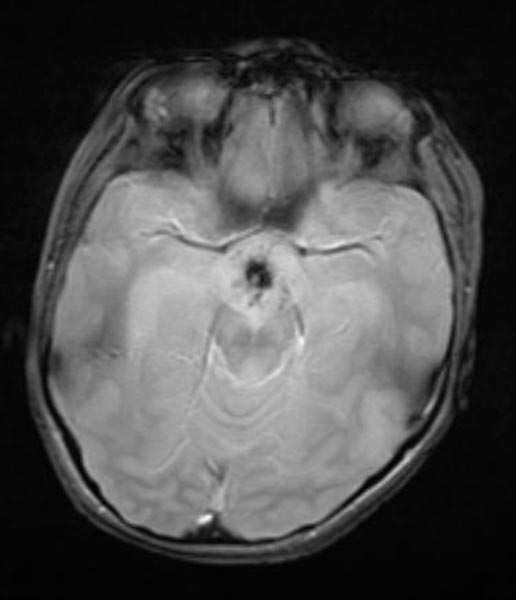
The findings on MRI are:
- Cysts
- Variable but 80% are mostly or partly T2 hyperintense
- T1: iso- to hyperintense to brain (due to high protein content machinery oil cysts)
- Solid component
- T1 C+ (Gd): vivid enhancement
- T2: variable or mixed
- Calcification
- Difficult to appreciate on conventional imaging
- Susceptible sequences may better demonstrate calcification
MR angiography: It may demonstrate displacement of the A1 segment of the anterior cerebral artery (ACA).
MR spectroscopy: Cyst contents may show a broad lipid spectrum, with an otherwise flat baseline.
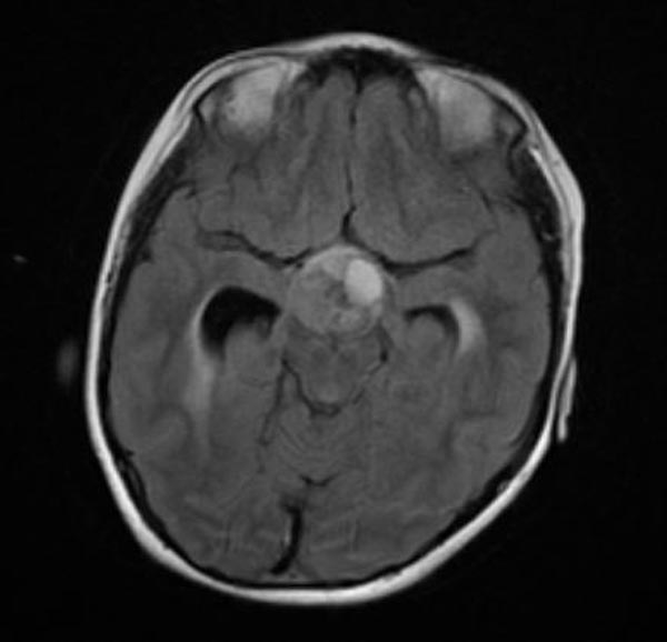
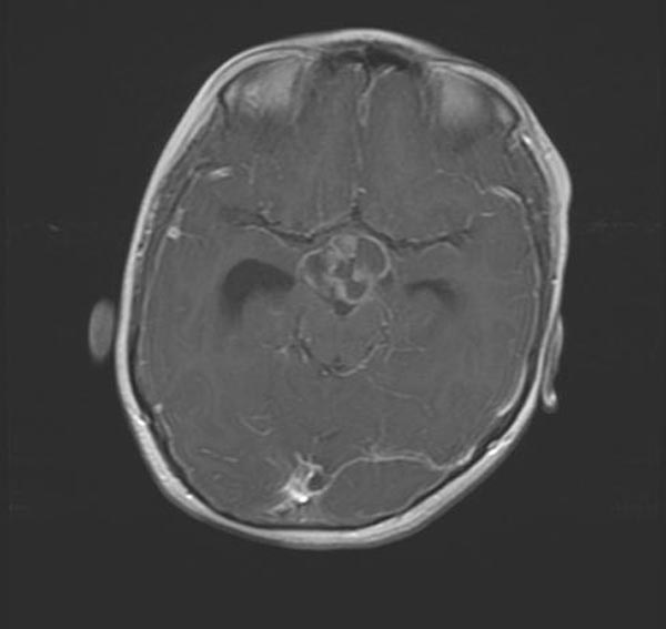
Papillary
The findings on MRI are:
- Cysts
- When present they are variable in signal
- 85% T1 hypointense
- Solid component
- T1: iso- to lightly hypointense to brain
- T1 C+: vivid enhancement
- T2: variable/mixed
MR spectroscopy: cyst contents does not show a broad lipid spectrum as they are filled with water fluid
-
Craniopharyngioma
-
Craniopharyngioma
-
Craniopharyngioma
-
Craniopharyngioma
-
Craniopharyngioma


