Craniopharyngioma MRI: Difference between revisions
Jump to navigation
Jump to search
No edit summary |
(→MRI) |
||
| Line 21: | Line 21: | ||
Image:Craniopharyngioma-005.jpg|Craniopharyngioma | Image:Craniopharyngioma-005.jpg|Craniopharyngioma | ||
</gallery> | </gallery> | ||
==References== | ==References== | ||
Revision as of 00:45, 23 August 2015
|
Craniopharyngioma Microchapters |
|
Diagnosis |
|---|
|
Treatment |
|
Case Studies |
|
Craniopharyngioma MRI On the Web |
|
American Roentgen Ray Society Images of Craniopharyngioma MRI |
Editor-In-Chief: C. Michael Gibson, M.S., M.D. [1]
Overview
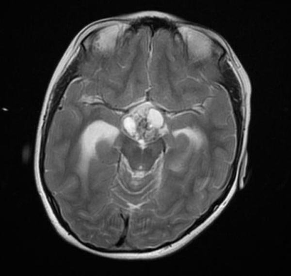
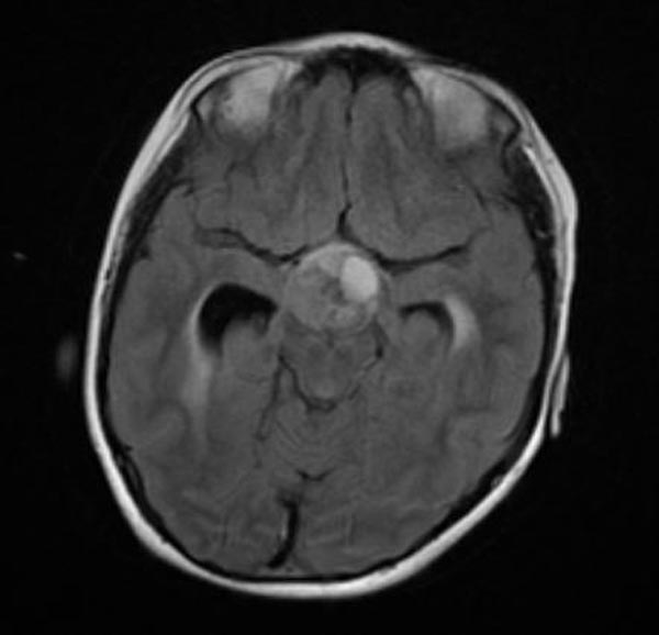
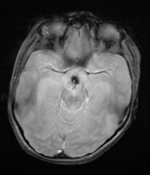
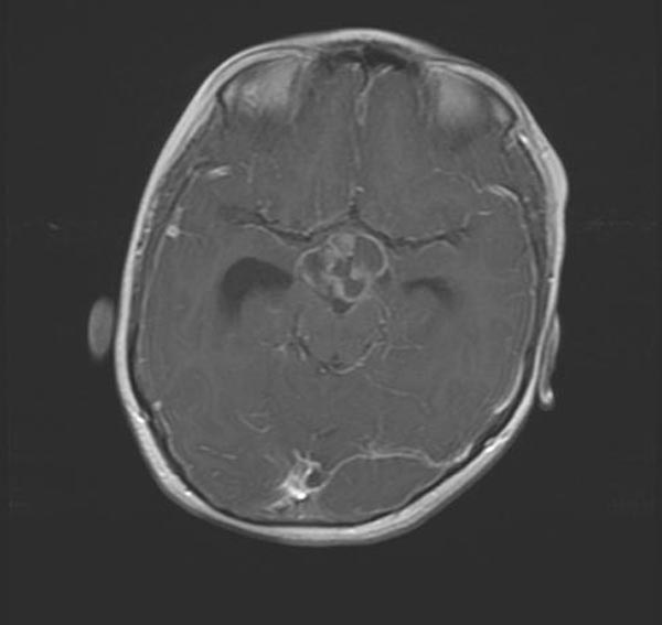
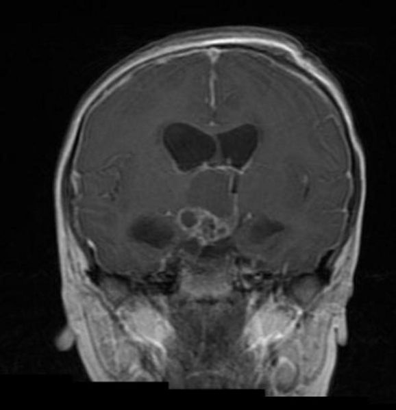
MRI
- Cranial magnetic resonance imaging (MRI): An MRI uses magnetic fields but it is a different type of image than what is produced by computed tomography (CT). It can produce very detailed images of the brain to help diagnose craniopharyngioma. Like computed tomography (CT), a contrast agent may be injected into a patient’s vein to create a better picture.
- Cranial computed tomography (CT) scan: CT scans are also used to diagnose craniopharyngioma. It can confirm the location of the tumor and show the organs nearby.
- Endocrine hormone tests
Adamantinomatous craniopharyngioma in a pediatric patient
-
Craniopharyngioma
-
Craniopharyngioma
-
Craniopharyngioma
-
Craniopharyngioma
-
Craniopharyngioma


