Colorado tick fever pathophysiology
|
Rocky Mountain spotted fever Microchapters |
|
Differentiating Rocky Mountain spotted fever from other Diseases |
|---|
|
Diagnosis |
|
Treatment |
|
Case Studies |
|
Colorado tick fever pathophysiology On the Web |
|
American Roentgen Ray Society Images of Colorado tick fever pathophysiology |
|
Directions to Hospitals Treating Rocky Mountain spotted fever |
|
Risk calculators and risk factors for Colorado tick fever pathophysiology |
Editor-In-Chief: C. Michael Gibson, M.S., M.D. [1] Associate Editor(s)-in-Chief: Ilan Dock, B.S.
Overview
Transmission
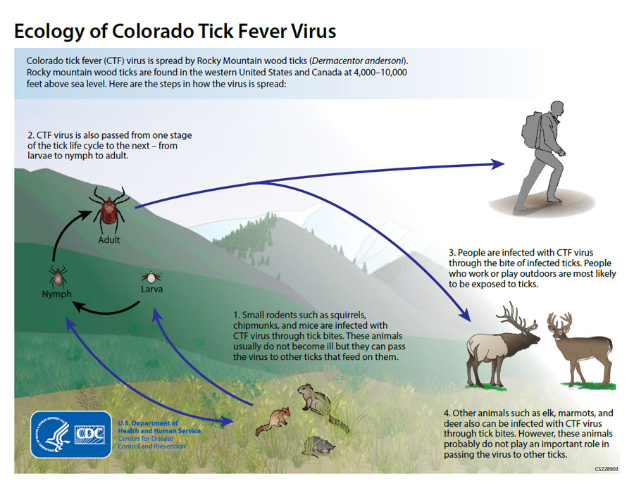
- Infection with Colorado tick fever occurs as a result of being bitten by an infected Rocky Mountain wood tick (Dermacentor andersoni).
- Colorado tick fever is transmitted to a tick during a blood meal involving a rodent reservoir such as a squirrels, chipmunks, and mice.
- Infection perpetuates as a tick continues to feed on another host.
- Viral transmission from human to human is rare, however may occur during blood transfusion.
