Bursitis overview: Difference between revisions
m (Bot: Removing from Primary care) |
|||
| (48 intermediate revisions by 5 users not shown) | |||
| Line 4: | Line 4: | ||
==Overview== | ==Overview== | ||
Bursitis is characterized by [[inflammation]] of a [[bursa]] and buildup of the fluid in the bursal sac. A [[bursa]] is a small, fluid-filled sac that acts as a cushion between a [[bone]] and other moving parts: [[muscles]], [[tendons]], or [[skin]]. Over 160 bursa are found throughout the [[body]] | Bursitis is characterized by the [[inflammation]] of a [[bursa]] and the buildup of the fluid in the bursal sac. A [[bursa]] is a small, fluid-filled sac that acts as a cushion between a [[bone]] and other moving parts: [[muscles]], [[tendons]], or [[skin]]. Over 160 bursa are found throughout the [[body]] but relatively few of them can cause bursitis. | ||
Based on the nature of inflammation bursitis may classified into 2 subtypes: [[septic]] and [[aseptic]]. | Based on the nature of the inflammation, bursitis may be classified into 2 subtypes: [[septic]] and [[aseptic]]. The most common bursitis subtypes include [[subacromial bursitis|subacromial]], [[olecranon bursitis|olecranon]], [[trochanteric bursitis|trochanteric]], [[prepatellar bursitis|prepatellar]], and [[retrocalcaneal bursitis|retrocalcaneal]]. | ||
Aseptic bursitis can be caused by | Aseptic bursitis can be caused by overuse or repetitive injury to the joint, abnormal and bony structure, and/or crystal deposit in the bursa. Additionally, septic bursitis can be caused by bacterial infection of the bursa through a skin injury following repetitive trauma. Common causes of septic bursitis include ''[[Staphylococcus aureus]]'', ''[[Staphylococcus epidermidis]]'', and ''[[Streptococcus|Streptococcus spp]]''. | ||
Bursitis must be differentiated from [[tendonitis]], [[cellulitis]], [[osteoarthritis]], [[ligament|ligamentous injuries]], and [[septic arthritis]]. | Bursitis must be differentiated from [[tendonitis]], [[cellulitis]], [[osteoarthritis]], [[ligament|ligamentous injuries]], and [[septic arthritis]]. | ||
The [[symptoms]] of bursitis differ based on the location of the [[inflammation]]. Focal swelling, pain, and redness are symptoms common to all forms of bursitis. | The [[symptoms]] of bursitis differ based on the location of the [[inflammation]]. Focal swelling, [[pain]], and redness are symptoms common to all forms of bursitis. | ||
The diagnosis of bursitis is usually made clinically. There are no diagnostic lab findings associated with bursitis. However, patients with septic bursitis may have elevated [[ESR]], [[CRP]], and [[white blood | The diagnosis of bursitis is usually made clinically. There are no diagnostic lab findings associated with bursitis. However, patients with septic bursitis may have elevated [[ESR]], [[CRP]], and [[white blood cell count]]. | ||
Ultrasonography may be a useful tool for confirming the diagnosis of bursitis, | [[Ultrasonography]] may be a useful tool for confirming the diagnosis of bursitis, while the aspiration of bursal fluids is usually reserved for the diagnosis of septic bursitis.<ref name="pmid9632407">{{cite journal| author=Stell IM, Gransden WR| title=Simple tests for septic bursitis: comparative study. | journal=BMJ | year= 1998 | volume= 316 | issue= 7148 | pages= 1877 | pmid=9632407 | doi= | pmc=28586 | url=https://www.ncbi.nlm.nih.gov/entrez/eutils/elink.fcgi?dbfrom=pubmed&tool=sumsearch.org/cite&retmode=ref&cmd=prlinks&id=9632407 }} </ref><ref name=Septic-olecranon-bursitis> Shell, Donald, Rob Perkins, and Andrew Cosgarea. "Septic olecranon bursitis: recognition and treatment." The Journal of the American Board of Family Practice 8.3 (1995): 217-220.</ref> | ||
Medical therapy for non-septic bursitis depends on the involved [[bursa]] and includes the RICE regimen (rest, ice, compression, elevation), NSAIDs, and/or [[corticosteroid]] injections. Restriction of activity is encouraged to prevent further injury and promote healing. Antimicrobials are the mainstay of therapy for septic bursitis. Surgical management is often reserved for non-responders.<ref name="pmid26577126">{{cite journal| author=Reilly D, Kamineni S| title=Olecranon bursitis. | journal=J Shoulder Elbow Surg | year= 2016 | volume= 25 | issue= 1 | pages= 158-67 | pmid=26577126 | doi=10.1016/j.jse.2015.08.032 | pmc= | url=https://www.ncbi.nlm.nih.gov/entrez/eutils/elink.fcgi?dbfrom=pubmed&tool=sumsearch.org/cite&retmode=ref&cmd=prlinks&id=26577126 }} </ref><ref name="pmid7667644">{{cite journal| author=Zimmermann B, Mikolich DJ, Ho G| title=Septic bursitis. | journal=Semin Arthritis Rheum | year= 1995 | volume= 24 | issue= 6 | pages= 391-410 | pmid=7667644 | doi= | pmc= | url=https://www.ncbi.nlm.nih.gov/entrez/eutils/elink.fcgi?dbfrom=pubmed&tool=sumsearch.org/cite&retmode=ref&cmd=prlinks&id=7667644 }} </ref><ref name="pmid21628647">{{cite journal| author=Aaron DL, Patel A, Kayiaros S, Calfee R| title=Four common types of bursitis: diagnosis and management. | journal=J Am Acad Orthop Surg | year= 2011 | volume= 19 | issue= 6 | pages= 359-67 | pmid=21628647 | doi= | pmc= | url=http://www.ncbi.nlm.nih.gov/entrez/eutils/elink.fcgi?dbfrom=pubmed&tool=sumsearch.org/cite&retmode=ref&cmd=prlinks&id=21628647 }} </ref> | Medical therapy for non-septic bursitis depends on the involved [[bursa]] and includes the [[RICE]] regimen (rest, ice, compression, elevation), [[NSAIDs]], and/or [[corticosteroid]] injections. Restriction of activity is encouraged to prevent further injury and promote healing. [[Antimicrobials]] are the mainstay of therapy for septic bursitis. Surgical management is often reserved for non-responders.<ref name="pmid26577126">{{cite journal| author=Reilly D, Kamineni S| title=Olecranon bursitis. | journal=J Shoulder Elbow Surg | year= 2016 | volume= 25 | issue= 1 | pages= 158-67 | pmid=26577126 | doi=10.1016/j.jse.2015.08.032 | pmc= | url=https://www.ncbi.nlm.nih.gov/entrez/eutils/elink.fcgi?dbfrom=pubmed&tool=sumsearch.org/cite&retmode=ref&cmd=prlinks&id=26577126 }} </ref><ref name="pmid7667644">{{cite journal| author=Zimmermann B, Mikolich DJ, Ho G| title=Septic bursitis. | journal=Semin Arthritis Rheum | year= 1995 | volume= 24 | issue= 6 | pages= 391-410 | pmid=7667644 | doi= | pmc= | url=https://www.ncbi.nlm.nih.gov/entrez/eutils/elink.fcgi?dbfrom=pubmed&tool=sumsearch.org/cite&retmode=ref&cmd=prlinks&id=7667644 }} </ref><ref name="pmid21628647">{{cite journal| author=Aaron DL, Patel A, Kayiaros S, Calfee R| title=Four common types of bursitis: diagnosis and management. | journal=J Am Acad Orthop Surg | year= 2011 | volume= 19 | issue= 6 | pages= 359-67 | pmid=21628647 | doi= | pmc= | url=http://www.ncbi.nlm.nih.gov/entrez/eutils/elink.fcgi?dbfrom=pubmed&tool=sumsearch.org/cite&retmode=ref&cmd=prlinks&id=21628647 }} </ref> | ||
===Classification=== | ===Classification=== | ||
Based on the nature of inflammation bursitis may classified into 2 subtypes: [[septic]] and [[aseptic]]. | Based on the nature of the inflammation, bursitis may classified into 2 subtypes: [[septic]] and [[aseptic]]. | ||
Common anatomic | Common anatomic locations of bursitis include the shoulder, elbow, hip, knee, and ankle. The most common bursitis subtypes include:<ref name="pmid22623812">{{cite journal| author=Chatra PS| title=Bursae around the knee joints. | journal=Indian J Radiol Imaging | year= 2012 | volume= 22 | issue= 1 | pages= 27-30 | pmid=22623812 | doi=10.4103/0971-3026.95400 | pmc=3354353 | url=http://www.ncbi.nlm.nih.gov/entrez/eutils/elink.fcgi?dbfrom=pubmed&tool=sumsearch.org/cite&retmode=ref&cmd=prlinks&id=22623812 }} </ref><ref name=Harrison-rheumatology> Fauci, Anthony S., and Carol Langford. Harrison's rheumatology. McGraw Hill Professional, 2010.</ref><ref name=bursitis-upper-limb> Walker‐Bone, Karen, et al. "Prevalence and impact of musculoskeletal disorders of the upper limb in the general population.</ref><ref name=Bursitis-four-types> Aaron, Daniel L., et al. "Four common types of bursitis: diagnosis and management." Journal of the American Academy of Orthopaedic Surgeons 19.6 (2011): 359-367.</ref> | ||
*[[Subacromial bursitis]] | *[[Subacromial bursitis]] | ||
*[[Olecranon bursitis]] | *[[Olecranon bursitis]] | ||
| Line 23: | Line 23: | ||
===Pathophysiology=== | ===Pathophysiology=== | ||
Bursitis is characterized by [[acute]] or [[chronic]] [[inflammation]] of a [[bursa]] and buildup of fluid in the bursa sac. A [[bursa]] is a small, fluid-filled sac that acts as a cushion between a [[bone]] and other moving parts: [[muscles]], [[tendons]], or [[skin]]. Over 160 | Bursitis is characterized by [[acute]] or [[chronic]] [[inflammation]] of a [[bursa]] and buildup of fluid in the bursa sac. A [[bursa]] is a small, fluid-filled sac that acts as a cushion between a [[bone]] and other moving parts: [[muscles]], [[tendons]], or [[skin]]. Over 160 bursae are found throughout the [[body]], though relatively few of them can cause bursitis. | ||
Aseptic bursitis can be caused by | Aseptic bursitis can be caused by overuse and repetitive injuries to the [[joint]], abnormal bony structure and crystal deposit in the bursa. It commonly affects the [[knee]] or [[elbow]] as a result of kneeling or leaning on the elbows longer than usual. Moreover, septic bursitis can be caused by bacterial infection of the bursa through the skin injury following repetitive trauma.<ref name=Harrison-rheumatology> Fauci, Anthony S., and Carol Langford. Harrison's rheumatology. McGraw Hill Professional, 2010.</ref><ref> Hellmann DB, Imboden JB., Jr. Musculoskeletal and immunologic disorders. In: McPhee SJ, Papadakis MA, editors. Current Medical Diagnosis & Treatment. McGraw-Hill Lange; 2010. pp. 2056–2061.</ref><ref name="pmid10090179">{{cite journal| author=García-Porrúa C, González-Gay MA, Ibañez D, García-País MJ| title=The clinical spectrum of severe septic bursitis in northwestern Spain: a 10 year study. | journal=J Rheumatol | year= 1999 | volume= 26 | issue= 3 | pages= 663-7 | pmid=10090179 | doi= | pmc= | url=https://www.ncbi.nlm.nih.gov/entrez/eutils/elink.fcgi?dbfrom=pubmed&tool=sumsearch.org/cite&retmode=ref&cmd=prlinks&id=10090179 }} </ref> | ||
====Images==== | ====Images==== | ||
The following | The following images depict different cases of bursitis:<ref name=Bursitis-pic> Wikimedia Commons. Bursitis. (2012) https://commons.wikimedia.org/wiki/Category:Bursitis Accessed on August 31, 2016</ref> | ||
<gallery>Image:Prepatellar bursitis.JPG|Prepatellar bursitis - By Thomas Kees - Own work (Original text: eigenes Archiv (selbst photographiert)), CC BY-SA 3.0 de, https://commons.wikimedia.org/w/index.php?curid=25418133 | |||
Image:Bursitispraepatellaris.jpg|Prepatellar bursitis - By Atropos235 - Own work, CC BY-SA 3.0, https://commons.wikimedia.org/w/index.php?curid=4079323 | |||
Image: | Image:Bursitis_Elbow_WC.JPG|Olecranon bursitis - By en:User:NJC123 - en:Image:Bursitis_Elbow_WC.JPG, Public Domain, https://commons.wikimedia.org/w/index.php?curid=2814450 | ||
</gallery> | </gallery> | ||
===Causes=== | ===Causes=== | ||
Common causes of bursitis include:<ref name="pmid9632407">{{cite journal| author=Stell IM, Gransden WR| title=Simple tests for septic bursitis: comparative study. | journal=BMJ | year= 1998 | volume= 316 | issue= 7148 | pages= 1877 | pmid=9632407 | doi= | pmc=28586 | url=https://www.ncbi.nlm.nih.gov/entrez/eutils/elink.fcgi?dbfrom=pubmed&tool=sumsearch.org/cite&retmode=ref&cmd=prlinks&id=9632407 }} </ref><ref name=bursitis-upper-limb> Walker‐Bone, Karen, et al. "Prevalence and impact of musculoskeletal disorders of the upper limb in the general population.</ref><ref name="pmid10987734">{{cite journal| author=Wang JP, Granlund KF, Bozzette SA, Botte MJ, Fierer J| title=Bursal sporotrichosis: case report and review. | journal=Clin Infect Dis | year= 2000 | volume= 31 | issue= 2 | pages= 615-6 | pmid=10987734 | doi=10.1086/313983 | pmc= | url=https://www.ncbi.nlm.nih.gov/entrez/eutils/elink.fcgi?dbfrom=pubmed&tool=sumsearch.org/cite&retmode=ref&cmd=prlinks&id=10987734 }} </ref><ref name=Bursitis-NIH> National Institute of Arthritis and Musculoskeletal and Skin disease, Bursitis. http://www.niams.nih.gov/Health_Info/Bursitis/default.asp Accessed August 25, 2016 </ref><ref name="pmid3074561">{{cite journal| author=McAfee JH, Smith DL| title=Olecranon and prepatellar bursitis. Diagnosis and treatment. | journal=West J Med | year= 1988 | volume= 149 | issue= 5 | pages= 607-10 | pmid=3074561 | doi= | pmc=1026560 | url=https://www.ncbi.nlm.nih.gov/entrez/eutils/elink.fcgi?dbfrom=pubmed&tool=sumsearch.org/cite&retmode=ref&cmd=prlinks&id=3074561 }} </ref> | |||
'''Aseptic bursitis:''' | |||
*Prolonged pressure, overuse, or strenuous activity | |||
**[[Elbow-joint|Elbow]]s and [[knee]]s are most commonly affected because they are rested upon more than many parts of the body with bursae and they also tend to endure the most repetitive use. | |||
**Shoulder bursitis is more commonly due to overuse of the shoulder joint and muscles. | |||
*Other inflammatory conditions (e.g., [[rheumatoid arthritis]] and [[spondyloarthritis]]) | |||
*[[Gout]] and [[pseudogout]] | |||
'''Septic bursitis:''' | |||
*''[[Staphylococcus aureus]]'' | |||
*''[[Staphylococcus epidermidis]]'' | |||
*''[[Streptococcus|Streptococcus spp]]'' | |||
===Differential Diagnosis=== | ===Differential Diagnosis=== | ||
| Line 40: | Line 51: | ||
===Epidemiology and Demographics=== | ===Epidemiology and Demographics=== | ||
Bursitis accounts for 400 visits per 100,000 visits to primary care clinic. The exact prevalence and incidence of bursitis | Bursitis accounts for 400 visits per 100,000 visits to primary care clinic. The exact prevalence and incidence of bursitis are unknown.<ref name="pmid3074561">{{cite journal| author=McAfee JH, Smith DL| title=Olecranon and prepatellar bursitis. Diagnosis and treatment. | journal=West J Med | year= 1988 | volume= 149 | issue= 5 | pages= 607-10 | pmid=3074561 | doi= | pmc=1026560 | url=https://www.ncbi.nlm.nih.gov/entrez/eutils/elink.fcgi?dbfrom=pubmed&tool=sumsearch.org/cite&retmode=ref&cmd=prlinks&id=3074561 }} </ref> | ||
===Risk Factors=== | ===Risk Factors=== | ||
Common risk factors in the development of bursitis include | Common risk factors in the development of bursitis include:<ref name="pmid3074561">{{cite journal| author=McAfee JH, Smith DL| title=Olecranon and prepatellar bursitis. Diagnosis and treatment. | journal=West J Med | year= 1988 | volume= 149 | issue= 5 | pages= 607-10 | pmid=3074561 | doi= | pmc=1026560 | url=https://www.ncbi.nlm.nih.gov/entrez/eutils/elink.fcgi?dbfrom=pubmed&tool=sumsearch.org/cite&retmode=ref&cmd=prlinks&id=3074561 }} </ref> | ||
*[[rheumatoid arthritis]] | |||
*[[osteoarthritis]] | |||
*[[gout]] or [[pseudogout]] | |||
*[[cellulitis]] | |||
*[[diabetes mellitus]] | |||
*use of systemic [[glucocorticoids]] | |||
*[[alcoholism]] | |||
*[[malignancy]] | |||
*[[leukopenia]] | |||
*having a hobby or job that involves repetitive motions (e.g., bicycling, playing baseball, gardening, setting tiles) | |||
===Screening=== | ===Screening=== | ||
| Line 49: | Line 70: | ||
===Natural History, Complications, and Prognosis=== | ===Natural History, Complications, and Prognosis=== | ||
Bursitis is often caused by | Bursitis is often caused by overuse and repetitive injuries to the [[joint]]. Symptoms of bursitis may develop rapidly within 2 to 3 days in an acute form. Patients with bursitis usually present with [[edema]], [[erythema]], and [[tenderness]] over the involved joint. In most cases, after an appropriate lifestyle adjustment, bursitis will gradually clear within a few days to weeks without any long-term consequences. If left untreated, acute bursitis may lead to chronic bursitis, which can result in cicatricial [[adhesions]], reduced mobility, and progressive pain. | ||
With treatment and activities adjustment, septic and aseptic bursitis are associated with excellent prognosis. | With proper treatment and an activities adjustment, septic and aseptic bursitis are associated with an excellent prognosis. | ||
==Diagnosis== | ==Diagnosis== | ||
===History=== | ===History=== | ||
Obtaining a complete history will be helpful in determining whether the bursitis is associated with any specific activities.<ref name=Harrison-rheumatology> Fauci, Anthony S., and Carol Langford. Harrison's rheumatology. McGraw Hill Professional, 2010.</ref><ref name=bursitis-upper-limb> Walker‐Bone, Karen, et al. "Prevalence and impact of musculoskeletal disorders of the upper limb in the general population.</ref><ref name=Bursitis-four-types> Aaron, Daniel L., et al. "Four common types of bursitis: diagnosis and management." Journal of the American Academy of Orthopaedic Surgeons 19.6 (2011): 359-367.</ref> | |||
===Symptoms and Physical Examination=== | ===Symptoms and Physical Examination=== | ||
| Line 65: | Line 86: | ||
| style="padding: 5px 5px; background: #DCDCDC;" | '''Subacromial bursitis'''<ref name=bursitis-upper-limb> Walker‐Bone, Karen, et al. "Prevalence and impact of musculoskeletal disorders of the upper limb in the general population.</ref> | | style="padding: 5px 5px; background: #DCDCDC;" | '''Subacromial bursitis'''<ref name=bursitis-upper-limb> Walker‐Bone, Karen, et al. "Prevalence and impact of musculoskeletal disorders of the upper limb in the general population.</ref> | ||
| style="padding: 5px 5px; background: #F5F5F5;" | | | style="padding: 5px 5px; background: #F5F5F5;" | | ||
*Mid shoulder pain | *Mid-shoulder pain | ||
* | *Pain often worse at night | ||
*Pain after repetitive activity (painting | *Pain after repetitive activity (e.g., painting, throwing a ball) | ||
*Popping sensation with shoulder movements | *Popping sensation with shoulder movements | ||
| style="padding: 5px 5px; background: #F5F5F5;" | | | style="padding: 5px 5px; background: #F5F5F5;" | | ||
*Redness, swelling, and warmth over the shoulder | *[[Redness]], [[swelling]], and warmth over the shoulder | ||
*Shoulder stiffness | *Shoulder stiffness | ||
*Lateral or anterior shoulder tenderness on palpation | *Lateral or anterior shoulder tenderness on palpation | ||
*Reduced active range of motion (ROM) with decreased elevation, internal rotation and abduction | *Reduced active [[range of motion]] (ROM) with decreased elevation, internal rotation, and abduction | ||
|- | |- | ||
| style="padding: 5px 5px; background: #DCDCDC;" | '''Olecranon bursitis'''<ref name="pmid8894865">{{cite journal| author=Stell IM| title=Septic and non-septic olecranon bursitis in the accident and emergency department--an approach to management. | journal=J Accid Emerg Med | year= 1996 | volume= 13 | issue= 5 | pages= 351-3 | pmid=8894865 | doi= | pmc=1342774 | url=https://www.ncbi.nlm.nih.gov/entrez/eutils/elink.fcgi?dbfrom=pubmed&tool=sumsearch.org/cite&retmode=ref&cmd=prlinks&id=8894865 }} </ref><ref name="pmid21075998">{{cite journal| author=Lockman L| title=Treating nonseptic olecranon bursitis: a 3-step technique. | journal=Can Fam Physician | year= 2010 | volume= 56 | issue= 11 | pages= 1157 | pmid=21075998 | doi= | pmc=2980436 | url=https://www.ncbi.nlm.nih.gov/entrez/eutils/elink.fcgi?dbfrom=pubmed&tool=sumsearch.org/cite&retmode=ref&cmd=prlinks&id=21075998 }} </ref> | | style="padding: 5px 5px; background: #DCDCDC;" | '''Olecranon bursitis'''<ref name="pmid8894865">{{cite journal| author=Stell IM| title=Septic and non-septic olecranon bursitis in the accident and emergency department--an approach to management. | journal=J Accid Emerg Med | year= 1996 | volume= 13 | issue= 5 | pages= 351-3 | pmid=8894865 | doi= | pmc=1342774 | url=https://www.ncbi.nlm.nih.gov/entrez/eutils/elink.fcgi?dbfrom=pubmed&tool=sumsearch.org/cite&retmode=ref&cmd=prlinks&id=8894865 }} </ref><ref name="pmid21075998">{{cite journal| author=Lockman L| title=Treating nonseptic olecranon bursitis: a 3-step technique. | journal=Can Fam Physician | year= 2010 | volume= 56 | issue= 11 | pages= 1157 | pmid=21075998 | doi= | pmc=2980436 | url=https://www.ncbi.nlm.nih.gov/entrez/eutils/elink.fcgi?dbfrom=pubmed&tool=sumsearch.org/cite&retmode=ref&cmd=prlinks&id=21075998 }} </ref> | ||
| Line 79: | Line 100: | ||
*Painful or painless focal swelling at the posterior elbow | *Painful or painless focal swelling at the posterior elbow | ||
| style="padding: 5px 5px; background: #F5F5F5;" | | | style="padding: 5px 5px; background: #F5F5F5;" | | ||
*Abrasion or contusion of skin (in a case of trauma) | *[[Abrasion]] or [[contusion]] of skin (in a case of trauma) | ||
*Swelling | *[[Swelling]] at the posterior elbow | ||
*Goose egg appearance over the olecranon process | *"Goose-egg" appearance over the [[olecranon process]] | ||
*Tenderness for palpation at the affected site | *[[Tenderness]] for palpation at the affected site | ||
*Systematic inflammatory processes | *Systematic [[inflammatory processes]] | ||
**Fever | **[[Fever]] | ||
**Rheumatoid nodules | **[[Rheumatoid nodules]] | ||
|- | |- | ||
| style="padding: 5px 5px; background: #DCDCDC;" | '''Trochanteric bursitis'''<ref name="pmid17880718">{{cite journal| author=Brinks A, van Rijn RM, Bohnen AM, Slee GL, Verhaar JA, Koes BW et al.| title=Effect of corticosteroid injection for trochanter pain syndrome: design of a randomised clinical trial in general practice. | journal=BMC Musculoskelet Disord | year= 2007 | volume= 8 | issue= | pages= 95 | pmid=17880718 | doi=10.1186/1471-2474-8-95 | pmc=2045096 | url=https://www.ncbi.nlm.nih.gov/entrez/eutils/elink.fcgi?dbfrom=pubmed&tool=sumsearch.org/cite&retmode=ref&cmd=prlinks&id=17880718 }} </ref><ref name="pmid4055877">{{cite journal| author=Karpinski MR, Piggott H| title=Greater trochanteric pain syndrome. A report of 15 cases. | journal=J Bone Joint Surg Br | year= 1985 | volume= 67 | issue= 5 | pages= 762-3 | pmid=4055877 | doi= | pmc= | url=https://www.ncbi.nlm.nih.gov/entrez/eutils/elink.fcgi?dbfrom=pubmed&tool=sumsearch.org/cite&retmode=ref&cmd=prlinks&id=4055877 }} </ref> | | style="padding: 5px 5px; background: #DCDCDC;" | '''Trochanteric bursitis'''<ref name="pmid17880718">{{cite journal| author=Brinks A, van Rijn RM, Bohnen AM, Slee GL, Verhaar JA, Koes BW et al.| title=Effect of corticosteroid injection for trochanter pain syndrome: design of a randomised clinical trial in general practice. | journal=BMC Musculoskelet Disord | year= 2007 | volume= 8 | issue= | pages= 95 | pmid=17880718 | doi=10.1186/1471-2474-8-95 | pmc=2045096 | url=https://www.ncbi.nlm.nih.gov/entrez/eutils/elink.fcgi?dbfrom=pubmed&tool=sumsearch.org/cite&retmode=ref&cmd=prlinks&id=17880718 }} </ref><ref name="pmid4055877">{{cite journal| author=Karpinski MR, Piggott H| title=Greater trochanteric pain syndrome. A report of 15 cases. | journal=J Bone Joint Surg Br | year= 1985 | volume= 67 | issue= 5 | pages= 762-3 | pmid=4055877 | doi= | pmc= | url=https://www.ncbi.nlm.nih.gov/entrez/eutils/elink.fcgi?dbfrom=pubmed&tool=sumsearch.org/cite&retmode=ref&cmd=prlinks&id=4055877 }} </ref> | ||
| style="padding: 5px 5px; background: #F5F5F5;" | | | style="padding: 5px 5px; background: #F5F5F5;" | | ||
*Pain in the lateral side of the hip | *Pain in the lateral side of the hip while walking, running or stair-climbing | ||
* | *Weakness of [[lower extremities]] | ||
*Pain with active and passive motion | *Pain with active and passive motion | ||
| style="padding: 5px 5px; background: #F5F5F5;" | | | style="padding: 5px 5px; background: #F5F5F5;" | | ||
*Tenderness at lateral hip, aggravated by active and passive external rotation and abduction | *[[Tenderness]] at lateral hip, aggravated by active and passive external rotation and abduction | ||
*Lateral hip pain on direct palpation | *Lateral hip pain on direct palpation | ||
*Weakness of the hip-abductors | *Weakness of the hip-abductors | ||
| Line 99: | Line 120: | ||
| style="padding: 5px 5px; background: #DCDCDC;" | '''Prepatellar bursitis'''<ref name=Bursitis-four-types> Aaron, Daniel L., et al. "Four common types of bursitis: diagnosis and management." Journal of the American Academy of Orthopaedic Surgeons 19.6 (2011): 359-367.</ref><ref name=Prepatellar-Bursitis> Huang, Yu-Chih, and Wen-Lin Yeh. "Endoscopic treatment of prepatellar bursitis." International orthopaedics 35.3 (2011): 355-358.</ref> | | style="padding: 5px 5px; background: #DCDCDC;" | '''Prepatellar bursitis'''<ref name=Bursitis-four-types> Aaron, Daniel L., et al. "Four common types of bursitis: diagnosis and management." Journal of the American Academy of Orthopaedic Surgeons 19.6 (2011): 359-367.</ref><ref name=Prepatellar-Bursitis> Huang, Yu-Chih, and Wen-Lin Yeh. "Endoscopic treatment of prepatellar bursitis." International orthopaedics 35.3 (2011): 355-358.</ref> | ||
| style="padding: 5px 5px; background: #F5F5F5;" | | | style="padding: 5px 5px; background: #F5F5F5;" | | ||
*Reduced range of motion at the knee | *Reduced [[range of motion]] at the knee | ||
*Focal swelling, pain, and redness | *Focal [[swelling]], [[pain]], and [[redness]] | ||
*Difficulty kneeling and walking | *Difficulty kneeling and walking | ||
| style="padding: 5px 5px; background: #F5F5F5;" | | | style="padding: 5px 5px; background: #F5F5F5;" | | ||
*Erythema at the affected site (knee) | *[[Erythema]] at the affected site (knee) | ||
*Ususally very large swelling over the knee | *Ususally very large swelling over the knee | ||
*Tenderness aggravated by bending and stretching the knee | *[[Tenderness]] aggravated by bending and stretching the knee | ||
*Reduced active range of motion (ROM) | *Reduced active [[range of motion]] (ROM) | ||
|- | |- | ||
| style="padding: 5px 5px; background: #DCDCDC;" | '''Retrocalcaneal bursitis'''<ref name=Harrison-rheumatology> Fauci, Anthony S., and Carol Langford. Harrison's rheumatology. McGraw Hill Professional, 2010.</ref><ref name=Achilles-tendon> Lyman, Jeffrey, Paul S. Weinhold, and Louis C. Almekinders. "Strain behavior of the distal Achilles tendon implications for insertional Achilles tendinopathy." The American Journal of Sports Medicine 32.2 (2004): 457-461.</ref> | | style="padding: 5px 5px; background: #DCDCDC;" | '''Retrocalcaneal bursitis'''<ref name=Harrison-rheumatology> Fauci, Anthony S., and Carol Langford. Harrison's rheumatology. McGraw Hill Professional, 2010.</ref><ref name=Achilles-tendon> Lyman, Jeffrey, Paul S. Weinhold, and Louis C. Almekinders. "Strain behavior of the distal Achilles tendon implications for insertional Achilles tendinopathy." The American Journal of Sports Medicine 32.2 (2004): 457-461.</ref> | ||
| style="padding: 5px 5px; background: #F5F5F5;" | | | style="padding: 5px 5px; background: #F5F5F5;" | | ||
*Swelling at the back of heel | *[[Swelling]] at the back of heel | ||
*Pain at the back of the heel, especially when running uphill | *[[Pain]] at the back of the heel, especially when running uphill | ||
*Pain while standing on tiptoes | *Pain while standing on tiptoes | ||
| style="padding: 5px 5px; background: #F5F5F5;" | | | style="padding: 5px 5px; background: #F5F5F5;" | | ||
*Swelling at the back of heel | *Swelling at the back of heel | ||
*Tenderness at the back of heel | *[[Tenderness]] at the back of heel | ||
*Painful ankle dorsiflexion | *Painful ankle [[dorsiflexion]] | ||
|} | |} | ||
===Laboratory Findings=== | ===Laboratory Findings=== | ||
The diagnosis of bursitis is usually made clinically. There are no diagnostic lab findings associated with bursitis. However, patients with septic bursitis may have elevated [[ESR]], [[CRP]], and [[white blood cells]].<ref name=Bursitis-Harrison's> Approach to Articular and Musculoskeletal Disorders, Harrison's Internal Medicine, 2011</ref><ref name= | The diagnosis of bursitis is usually made clinically. There are no diagnostic lab findings associated with bursitis. However, patients with septic bursitis may have elevated [[ESR]], [[CRP]], and [[white blood cells]].<ref name=Bursitis-Harrison's> Approach to Articular and Musculoskeletal Disorders, Harrison's Internal Medicine, 2011</ref><ref name=bursitis-upper-limb> Walker‐Bone, Karen, et al. "Prevalence and impact of musculoskeletal disorders of the upper limb in the general population.</ref><ref name=Bursitis-four-types> Aaron, Daniel L., et al. "Four common types of bursitis: diagnosis and management." Journal of the American Academy of Orthopaedic Surgeons 19.6 (2011): 359-367.</ref> | ||
===X ray=== | ===X ray=== | ||
X ray is | X ray is rarely required in patients with bursitis. | ||
X ray may be used as a diagnostic measure to support a clinical diagnosis of bursitis. | X ray may be used as a diagnostic measure to support a clinical diagnosis of bursitis. | ||
Joint x ray is generally reserved for patients with | Joint x ray is generally reserved for patients with histories of significant [[trauma]]. | ||
A | A standard x ray may be helpful in diagnosing a [[fracture]] or [[dislocation]].<ref name=Olecranon-Bursitis> Radiopedia. Olecranon Bursitis. http://radiopaedia.org/articles/olecranon-bursitis Accessed on August 23, 2016</ref><ref name=Prepatellar-Bursitis> Radiopedia. Prepatellar Bursitis. http://radiopaedia.org/cases/prepatellar-bursitis-1 Accessed on August 23, 2016</ref><ref name="pmid11519381">{{cite journal| author=Blankstein A, Cohen I, Diamant L, Heim M, Dudkiewicz I, Israeli A et al.| title=Achilles tendon pain and related pathologies: diagnosis by ultrasonography. | journal=Isr Med Assoc J | year= 2001 | volume= 3 | issue= 8 | pages= 575-8 | pmid=11519381 | doi= | pmc= | url=https://www.ncbi.nlm.nih.gov/entrez/eutils/elink.fcgi?dbfrom=pubmed&tool=sumsearch.org/cite&retmode=ref&cmd=prlinks&id=11519381 }} </ref> | ||
===CT=== | ===CT=== | ||
CT | CT scans are rarely required in patients with bursitis. CT scans are usually reserved for patients who do not respond to initial treatment. | ||
On CT scan, superficial bursitis may be characterized by fluid density at the subcutaneous tissue.<ref name=Olecranon-Bursitis> Radiopedia. Olecranon Bursitis. http://radiopaedia.org/articles/olecranon-bursitis Accessed on August 23, 2016</ref><ref name=Prepatellar-Bursitis> Radiopedia. Prepatellar Bursitis. http://radiopaedia.org/cases/prepatellar-bursitis-1 Accessed on August 23, 2016</ref> | On a CT scan, superficial bursitis may be characterized by fluid density at the [[subcutaneous tissue]].<ref name=Olecranon-Bursitis> Radiopedia. Olecranon Bursitis. http://radiopaedia.org/articles/olecranon-bursitis Accessed on August 23, 2016</ref><ref name=Prepatellar-Bursitis> Radiopedia. Prepatellar Bursitis. http://radiopaedia.org/cases/prepatellar-bursitis-1 Accessed on August 23, 2016</ref> | ||
===MRI=== | ===MRI=== | ||
MRI is | MRI is rarely required in patients with bursitis. Due to the associated cost and time requirements, the utility of MRI is limited compere to ultrasound. MRI is often reserved for patients who are likely to have other medical conditions such as [[tumors]], [[ligament|ligamentous injures]], or [[tendon|tendon injuries]]. | ||
On MRI, bursitis is characterized by bursal fluid collection, subcutaneous edema and joint effusion.<ref name=Olecranon-Bursitis> Radiopedia. Olecranon Bursitis. http://radiopaedia.org/articles/olecranon-bursitis Accessed on August 23, 2016</ref><ref name=Prepatellar-Bursitis> Radiopedia. Prepatellar Bursitis. http://radiopaedia.org/cases/prepatellar-bursitis-1 Accessed on August 23, 2016</ref> | On MRI, bursitis is characterized by bursal fluid collection, [[subcutaneous]] [[edema]], and joint effusion.<ref name=Olecranon-Bursitis> Radiopedia. Olecranon Bursitis. http://radiopaedia.org/articles/olecranon-bursitis Accessed on August 23, 2016</ref><ref name=Prepatellar-Bursitis> Radiopedia. Prepatellar Bursitis. http://radiopaedia.org/cases/prepatellar-bursitis-1 Accessed on August 23, 2016</ref> | ||
===Ultrasound=== | ===Ultrasound=== | ||
Ultrasonography may be a | [[Ultrasonography]] may be helpful in confirming a diagnosis of bursitis. On an ultrasound, bursitis may be characterized by bursal wall distention with presence of local hypoechoic or anechoic intra-bursal material, [[proliferation|synovial proliferation]], [[calcification]]s, and [[rheumatoid nodules]].<ref name=Bursitis-ultra-2> Blankstein A, Ganel A, Givon U, Mirovski Y, Chechick A. Ultrasonographic findings in patients with olecranon bursitis. Ultraschall Med 2006; 27: 568-571.</ref><ref name=Bursitis-ultra-1> Martinoli C, Bianchi S, Giovagnorio F, Pugliese F. Ultrasound of the elbow. Skeletal Radiol 2001; 30: 605-614</ref> | ||
===Other Diagnostic Studies=== | ===Other Diagnostic Studies=== | ||
Other diagnostic | Other diagnostic studies for bursitis include aspiration of the bursal fluid. | ||
Aspiration of bursal fluids is not recommended | Aspiration of bursal fluids is not recommended for the diagnosis of all types of bursitis. It is usually reserved for confirming a diagnosis of septic bursitis.<ref name="pmid9632407">{{cite journal| author=Stell IM, Gransden WR| title=Simple tests for septic bursitis: comparative study. | journal=BMJ | year= 1998 | volume= 316 | issue= 7148 | pages= 1877 | pmid=9632407 | doi= | pmc=28586 | url=https://www.ncbi.nlm.nih.gov/entrez/eutils/elink.fcgi?dbfrom=pubmed&tool=sumsearch.org/cite&retmode=ref&cmd=prlinks&id=9632407 }} </ref><ref name=Septic-olecranon-bursitis> Shell, Donald, Rob Perkins, and Andrew Cosgarea. "Septic olecranon bursitis: recognition and treatment." The Journal of the American Board of Family Practice 8.3 (1995): 217-220.</ref> | ||
==Treatment== | ==Treatment== | ||
===Medical Therapy=== | ===Medical Therapy=== | ||
Medical therapy for non-septic bursitis depends on the involved [[bursa]] and | Medical therapy for non-septic bursitis depends on the involved [[bursa]] and can include the [[RICE]] regimen (rest, ice, compression, elevation), [[NSAIDs]], and/or [[corticosteroid]] injections. Restriction of activity is encouraged to prevent further injury and promote healing. [[Antimicrobials]] are the mainstay of therapy for septic bursitis. Surgical intervention is generally reserved for non-responders.<ref name="pmid26577126">{{cite journal| author=Reilly D, Kamineni S| title=Olecranon bursitis. | journal=J Shoulder Elbow Surg | year= 2016 | volume= 25 | issue= 1 | pages= 158-67 | pmid=26577126 | doi=10.1016/j.jse.2015.08.032 | pmc= | url=https://www.ncbi.nlm.nih.gov/entrez/eutils/elink.fcgi?dbfrom=pubmed&tool=sumsearch.org/cite&retmode=ref&cmd=prlinks&id=26577126 }} </ref><ref name="pmid7667644">{{cite journal| author=Zimmermann B, Mikolich DJ, Ho G| title=Septic bursitis. | journal=Semin Arthritis Rheum | year= 1995 | volume= 24 | issue= 6 | pages= 391-410 | pmid=7667644 | doi= | pmc= | url=https://www.ncbi.nlm.nih.gov/entrez/eutils/elink.fcgi?dbfrom=pubmed&tool=sumsearch.org/cite&retmode=ref&cmd=prlinks&id=7667644 }} </ref><ref name="pmid21628647">{{cite journal| author=Aaron DL, Patel A, Kayiaros S, Calfee R| title=Four common types of bursitis: diagnosis and management. | journal=J Am Acad Orthop Surg | year= 2011 | volume= 19 | issue= 6 | pages= 359-67 | pmid=21628647 | doi= | pmc= | url=http://www.ncbi.nlm.nih.gov/entrez/eutils/elink.fcgi?dbfrom=pubmed&tool=sumsearch.org/cite&retmode=ref&cmd=prlinks&id=21628647 }} </ref> | ||
{| style="border: 0px; font-size: 90%; margin: 3px;" align=center | {| style="border: 0px; font-size: 90%; margin: 3px;" align=center | ||
| Line 152: | Line 177: | ||
*''[[Staphylococcus aureus]]'' bursitis often resolves with antibiotics alone | *''[[Staphylococcus aureus]]'' bursitis often resolves with antibiotics alone | ||
*''[[Sporothrix schenckii]]'' bursitis often requires [[bursectomy]] | *''[[Sporothrix schenckii]]'' bursitis often requires [[bursectomy]] | ||
*Most patients respond to oral antibiotics alone | *Most patients respond to oral antibiotics alone, though some require intravenous therapy | ||
| style="padding: 5px 5px; background: #F5F5F5" | | | style="padding: 5px 5px; background: #F5F5F5" | | ||
*Usually managed with RICE regimen (rest, ice, compression, elevation) | *Usually managed with [[RICE]] regimen (rest, ice, compression, elevation) | ||
*[[nonsteroidal anti-inflammatory drugs|Nonsteroidal anti-inflammatory drugs (NSAIDs]] | *[[nonsteroidal anti-inflammatory drugs|Nonsteroidal anti-inflammatory drugs (NSAIDs)]] | ||
*Local [[corticosteroid]] injections may be used in some patients who do not respond to initial therapy | *Local [[corticosteroid]] injections may be used in some patients who do not respond to initial therapy | ||
|} | |} | ||
===Surgery=== | ===Surgery=== | ||
[[Surgical intervention]] is not recommended for the management of bursitis. However, surgical techniques | [[Surgical intervention]] is not recommended for the management of bursitis. However, surgical techniques including [[bursectomy]] or longitudinal band release are usually reserved for patients with [[chronic]], recurrent, or [[septic]] bursitis.<ref name="pmid20521045">{{cite journal| author=Huang YC, Yeh WL| title=Endoscopic treatment of prepatellar bursitis. | journal=Int Orthop | year= 2011 | volume= 35 | issue= 3 | pages= 355-8 | pmid=20521045 | doi=10.1007/s00264-010-1033-5 | pmc=3047636 | url=https://www.ncbi.nlm.nih.gov/entrez/eutils/elink.fcgi?dbfrom=pubmed&tool=sumsearch.org/cite&retmode=ref&cmd=prlinks&id=20521045 }} </ref><ref name="pmid21814140">{{cite journal| author=Lustenberger DP, Ng VY, Best TM, Ellis TJ| title=Efficacy of treatment of trochanteric bursitis: a systematic review. | journal=Clin J Sport Med | year= 2011 | volume= 21 | issue= 5 | pages= 447-53 | pmid=21814140 | doi=10.1097/JSM.0b013e318221299c | pmc=3689218 | url=https://www.ncbi.nlm.nih.gov/entrez/eutils/elink.fcgi?dbfrom=pubmed&tool=sumsearch.org/cite&retmode=ref&cmd=prlinks&id=21814140 }} </ref> | ||
===Primary Prevention=== | ===Primary Prevention=== | ||
Effective measures for the primary prevention of bursitis include maintaining a healthy weight, taking breaks from repetitive tasks, using foam for | Effective measures for the primary prevention of bursitis include maintaining a healthy weight, taking regular breaks from repetitive tasks, using foam for knee- and elbow-pads, and practicing good posture.<ref name=Bursitis-prevention> National Institute of Arthritis and Musculoskeletal and Skin disease. Bursitis. http://www.niams.nih.gov/Health_Info/Bursitis/default.asp Accessed August 25, 2016</ref> | ||
===Secondary Prevention=== | ===Secondary Prevention=== | ||
There are no established | There are no established methods for secondary prevention of bursitis. However, a fast recovery may be facilitated by an adjustment in activities, the consistent use of foam for knee- or elbow-pads, and regular breaks during repetitive tasks.<ref name=Bursitis-prevention> National Institute of Arthritis and Musculoskeletal and Skin disease. Bursitis. http://www.niams.nih.gov/Health_Info/Bursitis/default.asp Accessed August 25, 2016</ref> | ||
==References== | ==References== | ||
{{reflist|2}} | {{reflist|2}} | ||
{{WS}} | |||
{{WH}} | |||
[[Category:Disease]] | |||
[[Category:Up-To-Date]] | |||
[[Category:Rheumatology]] | |||
[[Category:Orthopedics]] | [[Category:Orthopedics]] | ||
[[Category: | [[Category:Surgery]] | ||
[[Category: | [[Category:Emergency medicine]] | ||
[[Category:Infectious disease]] | [[Category:Infectious disease]] | ||
Latest revision as of 20:46, 29 July 2020
|
Bursitis Microchapters |
|
Diagnosis |
|---|
|
Treatment |
|
Case Studies |
|
Bursitis overview On the Web |
|
American Roentgen Ray Society Images of Bursitis overview |
Editor-In-Chief: C. Michael Gibson, M.S., M.D. [1]; Associate Editor(s)-in-Chief: Sara Mehrsefat, M.D. [2]
Overview
Bursitis is characterized by the inflammation of a bursa and the buildup of the fluid in the bursal sac. A bursa is a small, fluid-filled sac that acts as a cushion between a bone and other moving parts: muscles, tendons, or skin. Over 160 bursa are found throughout the body but relatively few of them can cause bursitis. Based on the nature of the inflammation, bursitis may be classified into 2 subtypes: septic and aseptic. The most common bursitis subtypes include subacromial, olecranon, trochanteric, prepatellar, and retrocalcaneal. Aseptic bursitis can be caused by overuse or repetitive injury to the joint, abnormal and bony structure, and/or crystal deposit in the bursa. Additionally, septic bursitis can be caused by bacterial infection of the bursa through a skin injury following repetitive trauma. Common causes of septic bursitis include Staphylococcus aureus, Staphylococcus epidermidis, and Streptococcus spp. Bursitis must be differentiated from tendonitis, cellulitis, osteoarthritis, ligamentous injuries, and septic arthritis. The symptoms of bursitis differ based on the location of the inflammation. Focal swelling, pain, and redness are symptoms common to all forms of bursitis. The diagnosis of bursitis is usually made clinically. There are no diagnostic lab findings associated with bursitis. However, patients with septic bursitis may have elevated ESR, CRP, and white blood cell count. Ultrasonography may be a useful tool for confirming the diagnosis of bursitis, while the aspiration of bursal fluids is usually reserved for the diagnosis of septic bursitis.[1][2] Medical therapy for non-septic bursitis depends on the involved bursa and includes the RICE regimen (rest, ice, compression, elevation), NSAIDs, and/or corticosteroid injections. Restriction of activity is encouraged to prevent further injury and promote healing. Antimicrobials are the mainstay of therapy for septic bursitis. Surgical management is often reserved for non-responders.[3][4][5]
Classification
Based on the nature of the inflammation, bursitis may classified into 2 subtypes: septic and aseptic. Common anatomic locations of bursitis include the shoulder, elbow, hip, knee, and ankle. The most common bursitis subtypes include:[6][7][8][9]
- Subacromial bursitis
- Olecranon bursitis
- Trochanteric bursitis
- Prepatellar bursitis
- Retrocalcaneal bursitis
Pathophysiology
Bursitis is characterized by acute or chronic inflammation of a bursa and buildup of fluid in the bursa sac. A bursa is a small, fluid-filled sac that acts as a cushion between a bone and other moving parts: muscles, tendons, or skin. Over 160 bursae are found throughout the body, though relatively few of them can cause bursitis. Aseptic bursitis can be caused by overuse and repetitive injuries to the joint, abnormal bony structure and crystal deposit in the bursa. It commonly affects the knee or elbow as a result of kneeling or leaning on the elbows longer than usual. Moreover, septic bursitis can be caused by bacterial infection of the bursa through the skin injury following repetitive trauma.[7][10][11]
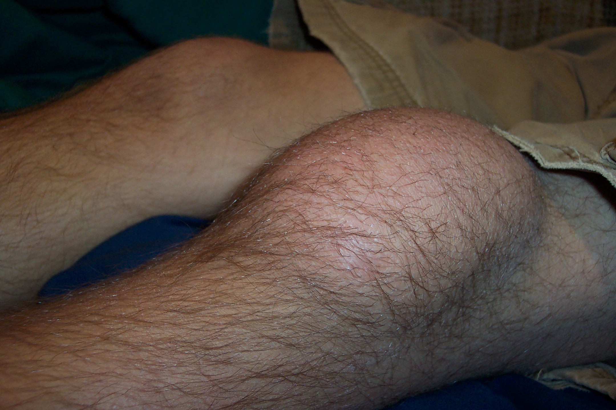
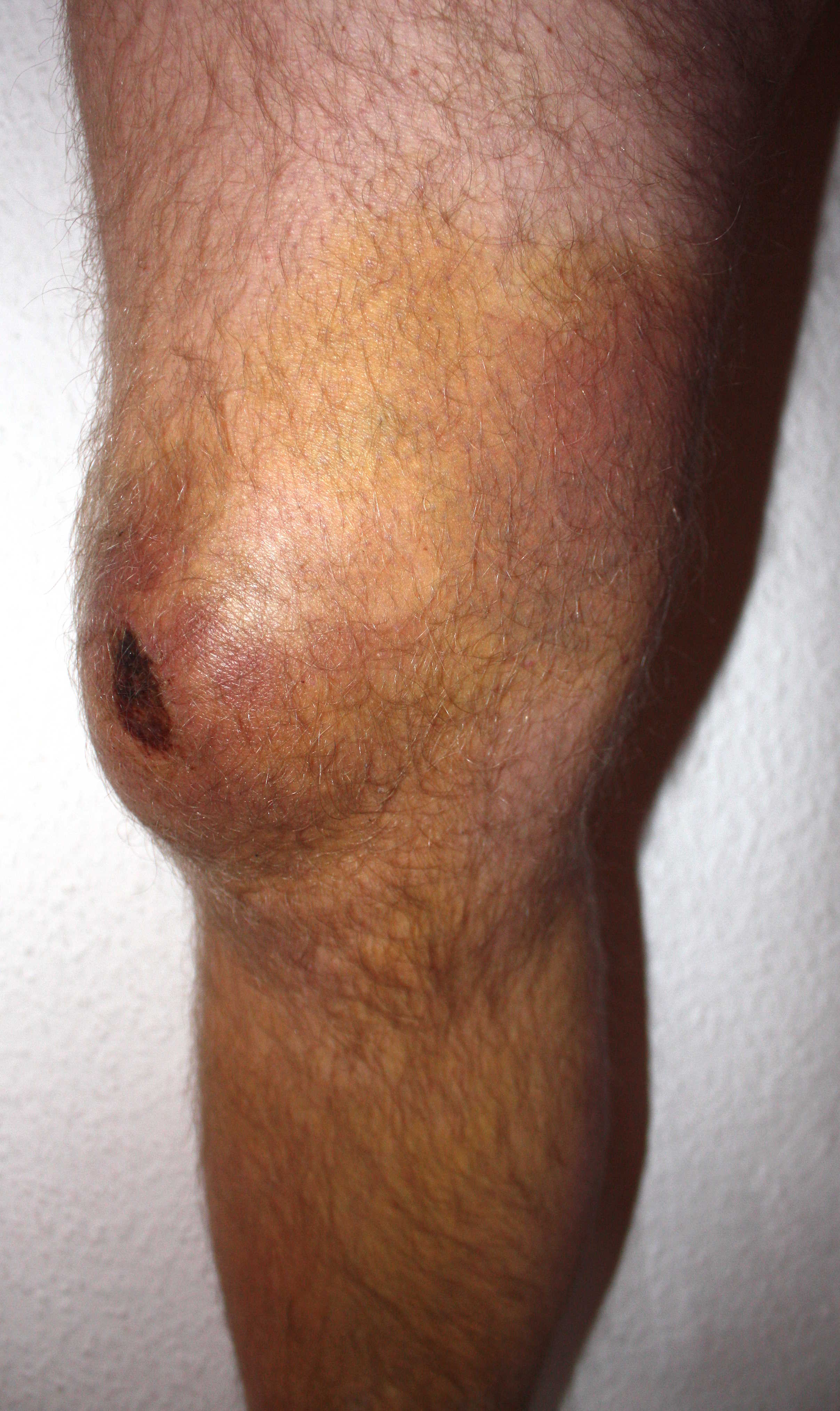
Images
The following images depict different cases of bursitis:[12]
-
Prepatellar bursitis - By Thomas Kees - Own work (Original text: eigenes Archiv (selbst photographiert)), CC BY-SA 3.0 de, https://commons.wikimedia.org/w/index.php?curid=25418133
-
Prepatellar bursitis - By Atropos235 - Own work, CC BY-SA 3.0, https://commons.wikimedia.org/w/index.php?curid=4079323
-
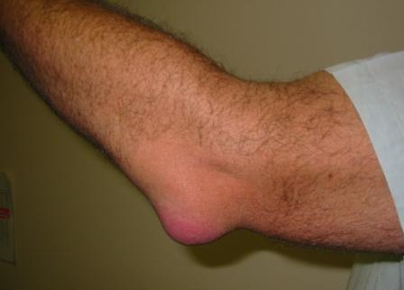
Olecranon bursitis - By en:User:NJC123 - en:Image:Bursitis_Elbow_WC.JPG, Public Domain, https://commons.wikimedia.org/w/index.php?curid=2814450


Causes
Common causes of bursitis include:[1][8][13][14][15]
Aseptic bursitis:
- Prolonged pressure, overuse, or strenuous activity
- Other inflammatory conditions (e.g., rheumatoid arthritis and spondyloarthritis)
- Gout and pseudogout
Septic bursitis:
Differential Diagnosis
Bursitis symptoms and signs are relatively non-specific. Even after detailed history and physical examination, imaging studies are often necessary to rule out other musculoskeletal conditions. Bursitis must be differentiated from tendonitis, cellulitis, osteoarthritis, ligamentous injuries, and septic arthritis.[7][8][9]
Epidemiology and Demographics
Bursitis accounts for 400 visits per 100,000 visits to primary care clinic. The exact prevalence and incidence of bursitis are unknown.[15]
Risk Factors
Common risk factors in the development of bursitis include:[15]
- rheumatoid arthritis
- osteoarthritis
- gout or pseudogout
- cellulitis
- diabetes mellitus
- use of systemic glucocorticoids
- alcoholism
- malignancy
- leukopenia
- having a hobby or job that involves repetitive motions (e.g., bicycling, playing baseball, gardening, setting tiles)
Screening
Screening for bursitis is not recommended.[16]
Natural History, Complications, and Prognosis
Bursitis is often caused by overuse and repetitive injuries to the joint. Symptoms of bursitis may develop rapidly within 2 to 3 days in an acute form. Patients with bursitis usually present with edema, erythema, and tenderness over the involved joint. In most cases, after an appropriate lifestyle adjustment, bursitis will gradually clear within a few days to weeks without any long-term consequences. If left untreated, acute bursitis may lead to chronic bursitis, which can result in cicatricial adhesions, reduced mobility, and progressive pain. With proper treatment and an activities adjustment, septic and aseptic bursitis are associated with an excellent prognosis.
Diagnosis
History
Obtaining a complete history will be helpful in determining whether the bursitis is associated with any specific activities.[7][8][9]
Symptoms and Physical Examination
| Type of Bursitis | Symptoms | Physical examination |
|---|---|---|
| Subacromial bursitis[8] |
|
|
| Olecranon bursitis[17][18] |
|
|
| Trochanteric bursitis[19][20] |
|
|
| Prepatellar bursitis[9][21] |
|
|
| Retrocalcaneal bursitis[7][22] |
|
Laboratory Findings
The diagnosis of bursitis is usually made clinically. There are no diagnostic lab findings associated with bursitis. However, patients with septic bursitis may have elevated ESR, CRP, and white blood cells.[23][8][9]
X ray
X ray is rarely required in patients with bursitis. X ray may be used as a diagnostic measure to support a clinical diagnosis of bursitis. Joint x ray is generally reserved for patients with histories of significant trauma. A standard x ray may be helpful in diagnosing a fracture or dislocation.[24][21][25]
CT
CT scans are rarely required in patients with bursitis. CT scans are usually reserved for patients who do not respond to initial treatment. On a CT scan, superficial bursitis may be characterized by fluid density at the subcutaneous tissue.[24][21]
MRI
MRI is rarely required in patients with bursitis. Due to the associated cost and time requirements, the utility of MRI is limited compere to ultrasound. MRI is often reserved for patients who are likely to have other medical conditions such as tumors, ligamentous injures, or tendon injuries. On MRI, bursitis is characterized by bursal fluid collection, subcutaneous edema, and joint effusion.[24][21]
Ultrasound
Ultrasonography may be helpful in confirming a diagnosis of bursitis. On an ultrasound, bursitis may be characterized by bursal wall distention with presence of local hypoechoic or anechoic intra-bursal material, synovial proliferation, calcifications, and rheumatoid nodules.[26][27]
Other Diagnostic Studies
Other diagnostic studies for bursitis include aspiration of the bursal fluid. Aspiration of bursal fluids is not recommended for the diagnosis of all types of bursitis. It is usually reserved for confirming a diagnosis of septic bursitis.[1][2]
Treatment
Medical Therapy
Medical therapy for non-septic bursitis depends on the involved bursa and can include the RICE regimen (rest, ice, compression, elevation), NSAIDs, and/or corticosteroid injections. Restriction of activity is encouraged to prevent further injury and promote healing. Antimicrobials are the mainstay of therapy for septic bursitis. Surgical intervention is generally reserved for non-responders.[3][4][5]
| Septic[3][4] | Aseptic |
|---|---|
|
|
Surgery
Surgical intervention is not recommended for the management of bursitis. However, surgical techniques including bursectomy or longitudinal band release are usually reserved for patients with chronic, recurrent, or septic bursitis.[28][29]
Primary Prevention
Effective measures for the primary prevention of bursitis include maintaining a healthy weight, taking regular breaks from repetitive tasks, using foam for knee- and elbow-pads, and practicing good posture.[30]
Secondary Prevention
There are no established methods for secondary prevention of bursitis. However, a fast recovery may be facilitated by an adjustment in activities, the consistent use of foam for knee- or elbow-pads, and regular breaks during repetitive tasks.[30]
References
- ↑ 1.0 1.1 1.2 Stell IM, Gransden WR (1998). "Simple tests for septic bursitis: comparative study". BMJ. 316 (7148): 1877. PMC 28586. PMID 9632407.
- ↑ 2.0 2.1 Shell, Donald, Rob Perkins, and Andrew Cosgarea. "Septic olecranon bursitis: recognition and treatment." The Journal of the American Board of Family Practice 8.3 (1995): 217-220.
- ↑ 3.0 3.1 3.2 Reilly D, Kamineni S (2016). "Olecranon bursitis". J Shoulder Elbow Surg. 25 (1): 158–67. doi:10.1016/j.jse.2015.08.032. PMID 26577126.
- ↑ 4.0 4.1 4.2 Zimmermann B, Mikolich DJ, Ho G (1995). "Septic bursitis". Semin Arthritis Rheum. 24 (6): 391–410. PMID 7667644.
- ↑ 5.0 5.1 Aaron DL, Patel A, Kayiaros S, Calfee R (2011). "Four common types of bursitis: diagnosis and management". J Am Acad Orthop Surg. 19 (6): 359–67. PMID 21628647.
- ↑ Chatra PS (2012). "Bursae around the knee joints". Indian J Radiol Imaging. 22 (1): 27–30. doi:10.4103/0971-3026.95400. PMC 3354353. PMID 22623812.
- ↑ 7.0 7.1 7.2 7.3 7.4 Fauci, Anthony S., and Carol Langford. Harrison's rheumatology. McGraw Hill Professional, 2010.
- ↑ 8.0 8.1 8.2 8.3 8.4 8.5 Walker‐Bone, Karen, et al. "Prevalence and impact of musculoskeletal disorders of the upper limb in the general population.
- ↑ 9.0 9.1 9.2 9.3 9.4 Aaron, Daniel L., et al. "Four common types of bursitis: diagnosis and management." Journal of the American Academy of Orthopaedic Surgeons 19.6 (2011): 359-367.
- ↑ Hellmann DB, Imboden JB., Jr. Musculoskeletal and immunologic disorders. In: McPhee SJ, Papadakis MA, editors. Current Medical Diagnosis & Treatment. McGraw-Hill Lange; 2010. pp. 2056–2061.
- ↑ García-Porrúa C, González-Gay MA, Ibañez D, García-País MJ (1999). "The clinical spectrum of severe septic bursitis in northwestern Spain: a 10 year study". J Rheumatol. 26 (3): 663–7. PMID 10090179.
- ↑ Wikimedia Commons. Bursitis. (2012) https://commons.wikimedia.org/wiki/Category:Bursitis Accessed on August 31, 2016
- ↑ Wang JP, Granlund KF, Bozzette SA, Botte MJ, Fierer J (2000). "Bursal sporotrichosis: case report and review". Clin Infect Dis. 31 (2): 615–6. doi:10.1086/313983. PMID 10987734.
- ↑ National Institute of Arthritis and Musculoskeletal and Skin disease, Bursitis. http://www.niams.nih.gov/Health_Info/Bursitis/default.asp Accessed August 25, 2016
- ↑ 15.0 15.1 15.2 McAfee JH, Smith DL (1988). "Olecranon and prepatellar bursitis. Diagnosis and treatment". West J Med. 149 (5): 607–10. PMC 1026560. PMID 3074561.
- ↑ U.S. Preventive Services Task Force http://www.uspreventiveservicestaskforce.org/BrowseRec/Search?s=bursitis Accessed on August 29, 2016
- ↑ Stell IM (1996). "Septic and non-septic olecranon bursitis in the accident and emergency department--an approach to management". J Accid Emerg Med. 13 (5): 351–3. PMC 1342774. PMID 8894865.
- ↑ Lockman L (2010). "Treating nonseptic olecranon bursitis: a 3-step technique". Can Fam Physician. 56 (11): 1157. PMC 2980436. PMID 21075998.
- ↑ Brinks A, van Rijn RM, Bohnen AM, Slee GL, Verhaar JA, Koes BW; et al. (2007). "Effect of corticosteroid injection for trochanter pain syndrome: design of a randomised clinical trial in general practice". BMC Musculoskelet Disord. 8: 95. doi:10.1186/1471-2474-8-95. PMC 2045096. PMID 17880718.
- ↑ Karpinski MR, Piggott H (1985). "Greater trochanteric pain syndrome. A report of 15 cases". J Bone Joint Surg Br. 67 (5): 762–3. PMID 4055877.
- ↑ 21.0 21.1 21.2 21.3 Huang, Yu-Chih, and Wen-Lin Yeh. "Endoscopic treatment of prepatellar bursitis." International orthopaedics 35.3 (2011): 355-358.
- ↑ Lyman, Jeffrey, Paul S. Weinhold, and Louis C. Almekinders. "Strain behavior of the distal Achilles tendon implications for insertional Achilles tendinopathy." The American Journal of Sports Medicine 32.2 (2004): 457-461.
- ↑ Approach to Articular and Musculoskeletal Disorders, Harrison's Internal Medicine, 2011
- ↑ 24.0 24.1 24.2 Radiopedia. Olecranon Bursitis. http://radiopaedia.org/articles/olecranon-bursitis Accessed on August 23, 2016
- ↑ Blankstein A, Cohen I, Diamant L, Heim M, Dudkiewicz I, Israeli A; et al. (2001). "Achilles tendon pain and related pathologies: diagnosis by ultrasonography". Isr Med Assoc J. 3 (8): 575–8. PMID 11519381.
- ↑ Blankstein A, Ganel A, Givon U, Mirovski Y, Chechick A. Ultrasonographic findings in patients with olecranon bursitis. Ultraschall Med 2006; 27: 568-571.
- ↑ Martinoli C, Bianchi S, Giovagnorio F, Pugliese F. Ultrasound of the elbow. Skeletal Radiol 2001; 30: 605-614
- ↑ Huang YC, Yeh WL (2011). "Endoscopic treatment of prepatellar bursitis". Int Orthop. 35 (3): 355–8. doi:10.1007/s00264-010-1033-5. PMC 3047636. PMID 20521045.
- ↑ Lustenberger DP, Ng VY, Best TM, Ellis TJ (2011). "Efficacy of treatment of trochanteric bursitis: a systematic review". Clin J Sport Med. 21 (5): 447–53. doi:10.1097/JSM.0b013e318221299c. PMC 3689218. PMID 21814140.
- ↑ 30.0 30.1 National Institute of Arthritis and Musculoskeletal and Skin disease. Bursitis. http://www.niams.nih.gov/Health_Info/Bursitis/default.asp Accessed August 25, 2016