Bicuspid aortic stenosis chest x ray

Jump to navigation Jump to search
The printable version is no longer supported and may have rendering errors. Please update your browser bookmarks and please use the default browser print function instead.

Bicuspid aortic stenosis Microchapters

Home

Patient Info

Overview

Anatomy

Historical Perspective

Pathophysiology

Epidemiology & Demographics

Risk Factors

Screening

Causes of Bicuspid aortic stenosis

Natural History, Complications & Prognosis

Diagnosis

History & Symptoms

Physical Examination

Lab Tests

Electrocardiogram

Chest X Ray

CT

MRI

Echocardiography

Cardiac Catheterization

Treatment

Overview

Medical Therapy

Surgery

Bicuspid aortic stenosis chest x ray On the Web

Most recent articles

Most cited articles

Review articles

CME Programs

Powerpoint slides

Images

American Roentgen Ray Society Images of Bicuspid aortic stenosis chest x ray

All Images
X-rays
Echo & Ultrasound
CT Images
MRI

Ongoing Trials at Clinical Trials.gov

US National Guidelines Clearinghouse

NICE Guidance

FDA on Bicuspid aortic stenosis chest x ray

CDC on Bicuspid aortic stenosis chest x ray

Bicuspid aortic stenosis chest x ray in the news

Blogs on Bicuspid aortic stenosis chest x ray

Directions to Hospitals Treating Bicuspid aortic stenosis

Risk calculators and risk factors for Bicuspid aortic stenosis chest x ray

Editor-In-Chief: C. Michael Gibson, M.S., M.D. [1]; Associate Editor(s)-In-Chief: Varun Kumar, M.B.B.S. [2]; Usama Talib, BSc, MD [3]

Overview

Chest x ray may be used as a diagnostic tool in the evaluation of aortic stenosis. Findings associated with aortic stenosis include left ventricular hypertrophy.

Chest X Ray
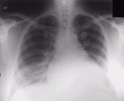
Chest x-ray shows left ventricular Hypertrophy due to aortic stenosis

Chest X-ray may show hypertrophied left ventricle if there is aortic stenosis as shown here. In later stages of disease; the left ventricle dilates and the patient may have pulmonary congestion which may be appearant on X-ray. In case of severe aortic stenosis for a long time; the left atrium, pulmonary artery, and right side of heart may become enlarged too.
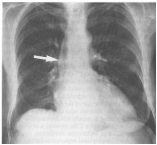
Chest x-ray shows prominent aorta with post-stenotic dilatation of the aortic valve.


References

Template:WS Template:WH CME Category::Cardiology