Atelectasis chest x ray
|
Atelectasis Microchapters |
|
Diagnosis |
|---|
|
Treatment |
|
Case Studies |
|
Atelectasis chest x ray On the Web |
|
American Roentgen Ray Society Images of Atelectasis chest x ray |
|
Risk calculators and risk factors for Atelectasis chest x ray |
Please help WikiDoc by adding content here. It's easy! Click here to learn about editing.
Editor-In-Chief: C. Michael Gibson, M.S., M.D. [1]
Chest X Ray
Post-surgical atelectasis will be bibasal in pattern.
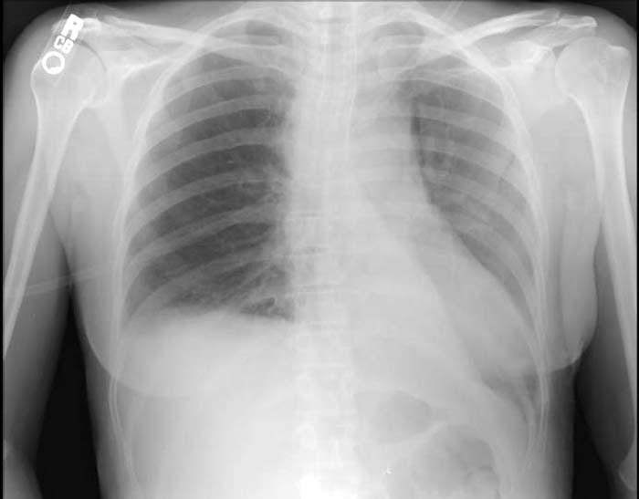
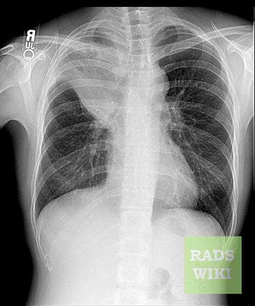
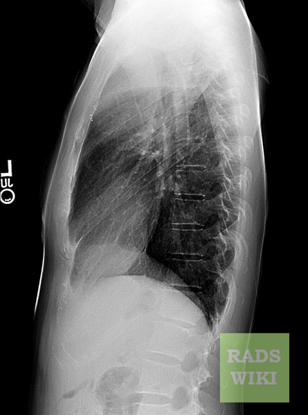
Images shown in this section are courtesy of RadsWiki and copylefted.
-
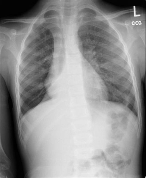
Right lower lobe collapse
-
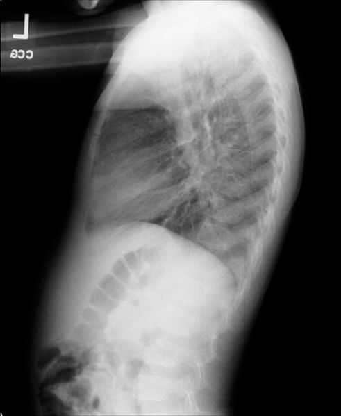
Right lower lobe collapse. The same patient. Lateral view.


-
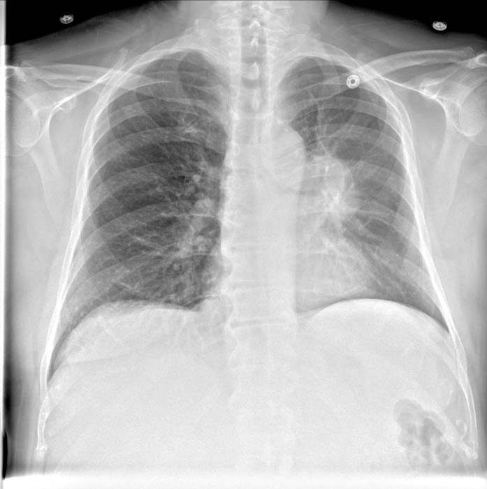
Left upper lobe collapse
-
Left upper lobe collapse


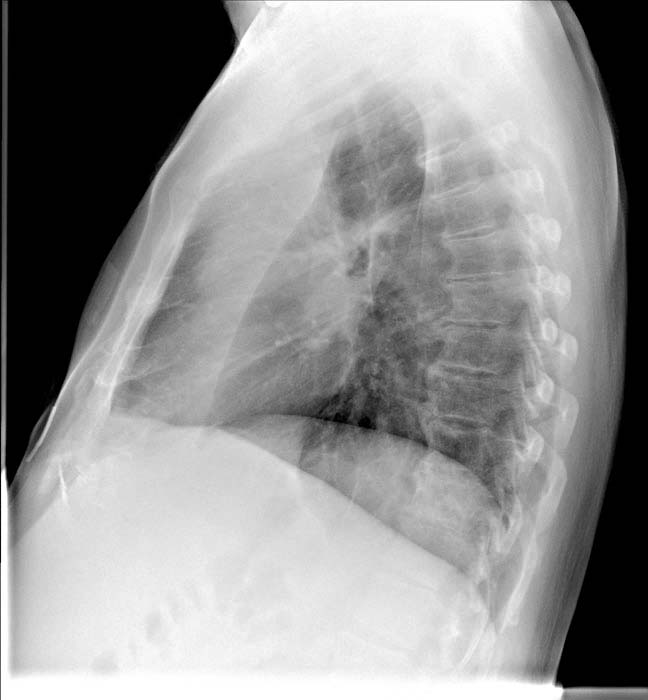
-
Left lower lobe collapse

-
S Sign of the Golden
-
S Sign of the Golden

