Atelectasis chest x ray: Difference between revisions
(→X Ray) |
No edit summary |
||
| Line 4: | Line 4: | ||
{{CMG}}; {{AE}} | {{CMG}}; {{AE}} | ||
==Overview== | ==Overview== | ||
An x-ray may be helpful in the diagnosis of atelectasis. Findings on an x-ray suggestive of atelectasis include [[displacement]] of [[Fissure|fissures]], [[rib]] crowding, elevation of ipsilateral [[Thoracic diaphragm|diaphragm]], volume loss on ipsilateral hemithorax, [[Hilum|hilar]] displacement and compensatory hyperlucency of the remaining lobes. Complete lung atelectasis and atelectasis involving different parts of the lung have their own characteristic appearance. While complete atelectasis of the lung may lead to opacification of the entire hemithorax and ipsilateral shift of the [[mediastinum]], a right midle and lower lobe atelectasis may show [[Pleural effusions|subpulmonic effusions]] along with right [[Thoracic diaphragm|hemidiaphragmatic]] elevation on X-ray. | |||
An x-ray may be helpful in the diagnosis of | |||
==X Ray== | ==X Ray== | ||
*An x-ray may be helpful in the diagnosis of atelectasis. Findings on an x-ray suggestive of atelectasis include: | |||
**Signs of lobar collapse such as: | |||
***[[Mediastinal|Shifting of the mediastinum]] towards the collapsed [[Lung|lung lobe]] | |||
***[[Hilum|Hilar]] displacement | |||
***Silhouetting of the [[Thoracic diaphragm|diaphragm]] or the heart border | |||
***[[Rib]] crowding | |||
***Compensatory hyperlucency of the remaining lobes | |||
***Elevation of [[Diaphragm|ipsilateral diaphragm]] | |||
***Opacification of the collapsed lung lobe | |||
***Displacement of fissures | |||
***Volume loss on ipsilateral hemithorax | |||
* | *X-ray findings in cases with complete atelectasis of the lung include: | ||
**Opacification of the entire hemithorax due to complete collapse of a lung | |||
* | **Ipsilateral shift of the [[mediastinum]], that helps distinguish atelectasis from [[pleural effusion]] | ||
**[ | *X-ray findings suggestive of right upper lobe (RUL) collapse include: | ||
**[ | **Medial and superior shift of RUL | ||
**[ | **Sign of Golden S: Concave appearance of minor [[fissure]] | ||
**Right minor [[fissure]] elevation | |||
* | **Right [[Hilum|hilar]] elevation | ||
** | *X-ray appearance of right middle lobe collapse: | ||
**[ | **Traingular opacity | ||
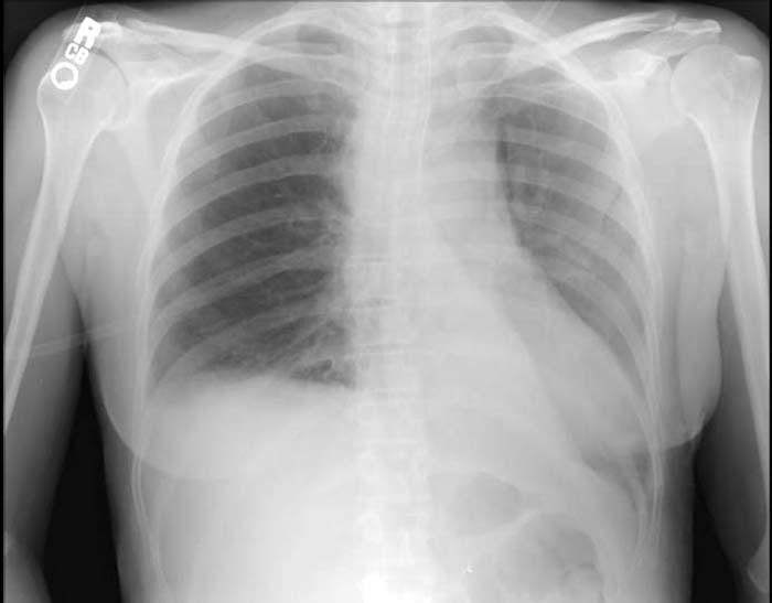
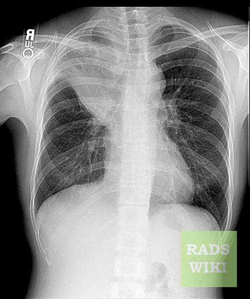
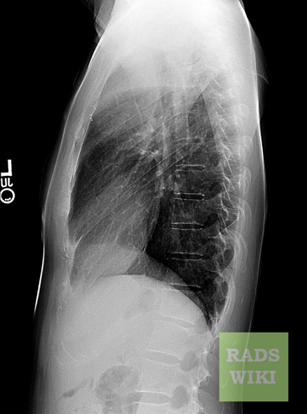
** | *X-ray appearance of right lower lobe (RLL) collapse: | ||
**Posterior and inferior shift of RLL due to collapse | |||
**Superior triangle sign: Rightward shift of structures in the superior [[mediastinum]] | |||
**Blurring of the right hemidiaphragm (posterior third) | |||
**Visibility of the major [[fissure]], which is usually not seen | |||
*X-ray appearance of a right midle and lower lobe atelectasis: | |||
**[[Pleural effusion|Subpulmonic effusion]] | |||
**Elevation of the right hemidiaphragm | |||
*X-ray appearance of left upper lobe (LUL) collapse: | |||
**Atelectatic left upper lobe shifts anteriorly and superiorly | |||
**PA view: Faint opacity of the atelectatic lobe in the left upper hemithorax | |||
* X-ray appearance of left lower lobe (LLL) collapse: | |||
** Retrocardiac opacity | |||
** Downward displacement of the [[hilum]] | |||
** Aortic-knob sign: Obliteration of the [[aortic arch]] by the [[superior mediastinum]] | |||
** Lateral view: Indistinct appearance of the posterior third of the [[Thoracic diaphragm|diaphragm]] due to opacity | |||
Retrocardiac opacity | |||
Downward displacement of the hilum | |||
Aortic-knob sign: | |||
Lateral | |||
* X-ray appearance of rounded atelectasis: | |||
** Subpleural mass | |||
** Location of rounded atelectasis: Right middle lobe, lower lobes or [[lingula]] | |||
** Comet-tail sign: Bronchovascular structures projecting out of the mass toward the [[hilum]], in a swirl appearance | |||
** [[Pleural plaque|Parietal pleural plaque]] | |||
* X-ray appearance of post-surgical atelectasis: | |||
* Bibasal pattern | |||
Images shown in this section are courtesy of RadsWiki and copylefted. | Images shown in this section are courtesy of RadsWiki and copylefted. | ||
| Line 95: | Line 62: | ||
</gallery> | </gallery> | ||
</div> | </div> | ||
<div align="left"> | <div align="left"> | ||
| Line 104: | Line 69: | ||
</gallery> | </gallery> | ||
</div> | </div> | ||
<div align="left"> | <div align="left"> | ||
| Line 112: | Line 75: | ||
</gallery> | </gallery> | ||
</div> | </div> | ||
<div align="left"> | <div align="left"> | ||
Revision as of 14:15, 22 February 2018
|
Atelectasis Microchapters |
|
Diagnosis |
|---|
|
Treatment |
|
Case Studies |
|
Atelectasis chest x ray On the Web |
|
American Roentgen Ray Society Images of Atelectasis chest x ray |
|
Risk calculators and risk factors for Atelectasis chest x ray |
Editor-In-Chief: C. Michael Gibson, M.S., M.D. [1]; Associate Editor(s)-in-Chief:
Overview
An x-ray may be helpful in the diagnosis of atelectasis. Findings on an x-ray suggestive of atelectasis include displacement of fissures, rib crowding, elevation of ipsilateral diaphragm, volume loss on ipsilateral hemithorax, hilar displacement and compensatory hyperlucency of the remaining lobes. Complete lung atelectasis and atelectasis involving different parts of the lung have their own characteristic appearance. While complete atelectasis of the lung may lead to opacification of the entire hemithorax and ipsilateral shift of the mediastinum, a right midle and lower lobe atelectasis may show subpulmonic effusions along with right hemidiaphragmatic elevation on X-ray.
X Ray
- An x-ray may be helpful in the diagnosis of atelectasis. Findings on an x-ray suggestive of atelectasis include:
- Signs of lobar collapse such as:
- Shifting of the mediastinum towards the collapsed lung lobe
- Hilar displacement
- Silhouetting of the diaphragm or the heart border
- Rib crowding
- Compensatory hyperlucency of the remaining lobes
- Elevation of ipsilateral diaphragm
- Opacification of the collapsed lung lobe
- Displacement of fissures
- Volume loss on ipsilateral hemithorax
- Signs of lobar collapse such as:
- X-ray findings in cases with complete atelectasis of the lung include:
- Opacification of the entire hemithorax due to complete collapse of a lung
- Ipsilateral shift of the mediastinum, that helps distinguish atelectasis from pleural effusion
- X-ray findings suggestive of right upper lobe (RUL) collapse include:
- X-ray appearance of right middle lobe collapse:
- Traingular opacity
- X-ray appearance of right lower lobe (RLL) collapse:
- Posterior and inferior shift of RLL due to collapse
- Superior triangle sign: Rightward shift of structures in the superior mediastinum
- Blurring of the right hemidiaphragm (posterior third)
- Visibility of the major fissure, which is usually not seen
- X-ray appearance of a right midle and lower lobe atelectasis:
- Subpulmonic effusion
- Elevation of the right hemidiaphragm
- X-ray appearance of left upper lobe (LUL) collapse:
- Atelectatic left upper lobe shifts anteriorly and superiorly
- PA view: Faint opacity of the atelectatic lobe in the left upper hemithorax
- X-ray appearance of left lower lobe (LLL) collapse:
- Retrocardiac opacity
- Downward displacement of the hilum
- Aortic-knob sign: Obliteration of the aortic arch by the superior mediastinum
- Lateral view: Indistinct appearance of the posterior third of the diaphragm due to opacity
- X-ray appearance of rounded atelectasis:
- Subpleural mass
- Location of rounded atelectasis: Right middle lobe, lower lobes or lingula
- Comet-tail sign: Bronchovascular structures projecting out of the mass toward the hilum, in a swirl appearance
- Parietal pleural plaque
- X-ray appearance of post-surgical atelectasis:
- Bibasal pattern
Images shown in this section are courtesy of RadsWiki and copylefted.
-
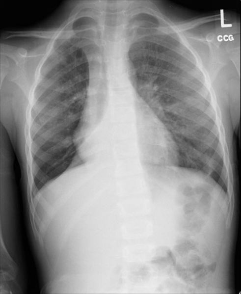
Right lower lobe collapse
-
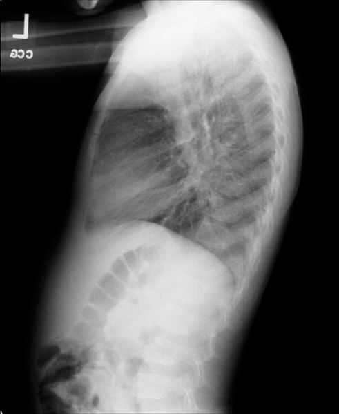
Right lower lobe collapse. The same patient. Lateral view.


-
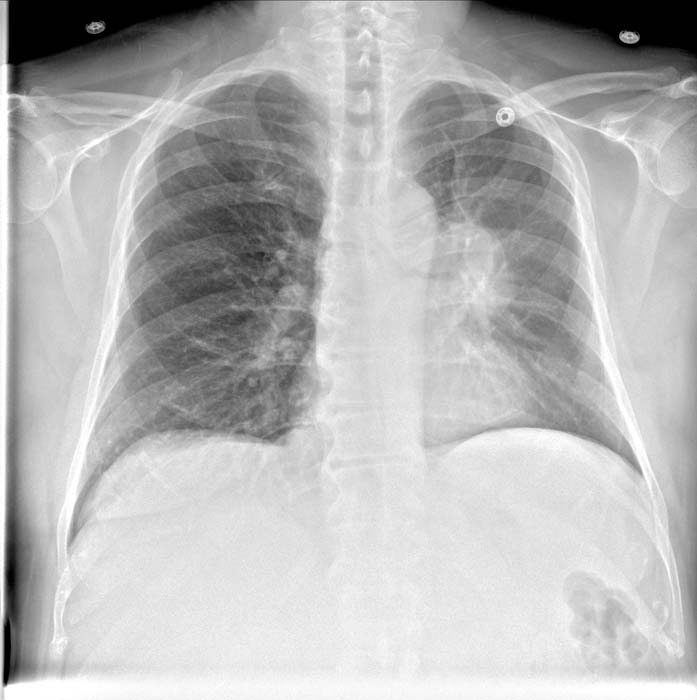
Left upper lobe collapse
-
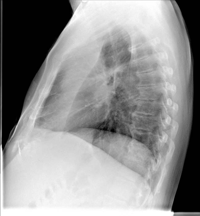
Left upper lobe collapse


-
Left lower lobe collapse

-
S Sign of the Golden
-
S Sign of the Golden

