Atelectasis CT: Difference between revisions
(→CT) |
No edit summary |
||
| Line 3: | Line 3: | ||
{{CMG}} {{AE}}{{Cherry}} | {{CMG}} {{AE}}{{Cherry}} | ||
==Overview== | ==Overview== | ||
CT findings suggestive of atelectasis include [[Hilum|hilar]] displacement, elevation of ipsilateral [[Thoracic diaphragm|diaphragm]], rib crowding, [[displacement]] of [[Fissure|fissures]], and compensatory hyperlucency of the remaining [[Lung|lobes]]. CT findings associated with complete atelectasis of an entire [[lung]] include opacification of the entire hemithorax and ipsilateral shift of the [[mediastinum]]. Collapse of different parts of the lung have their own characteristic appearance. For example, a collapsed right middle lobe has a tilted icecream sign which appears as a triangular [[opacity]] against the border of the right heart with a laterally pointed apex. On the other hand, RUL collapse appears as a right paratracheal opacity, with a concave lateral appearance of the [[Fissure|minor lung fissure]]. | |||
[ | |||
==CT== | ==CT== | ||
* | *CT scan may be helpful in the diagnosis of atelectasis. Findings on CT scan suggestive of atelectasis include direct and indirect signs of [[Collapse|lobar collapse]]: | ||
**Loss of volume on ipsilateral hemithorax | |||
**[[Rib]] crowding | |||
** | **Silhouetting of the [[Thoracic diaphragm|diaphragm]] or the heart border | ||
**[ | **Elevation of ipsilateral [[Thoracic diaphragm|diaphragm]] | ||
**[ | **[[Mediastinum|Mediastinal]] shift toward the side of collapse | ||
**[[Hilum|Hilar]] displacement | |||
**Opacification of the [[Collapsed lung|collapsed lobe]] | |||
[ | **[[Displacement]] of [[Fissure|fissures]] | ||
[ | **Compensatory hyperlucency of the remaining [[Lung|lobes]] | ||
*CT findings associated with complete atelectasis of an entire lung: | |||
**Complete collapse of a lung leads to opacification of the entire hemithorax | |||
**Ipsilateral shift of the [[mediastinum]], distinguishing atelectasis from massive [[pleural effusion]] | |||
Opacification of the collapsed lobe | |||
Compensatory hyperlucency of the remaining lobes | |||
Complete collapse of a lung leads to opacification of the entire hemithorax | |||
RUL collapse: | * CT findings associated with right upper lobe (RUL) collapse: | ||
RUL collapse appears as a right paratracheal opacity | ** RUL collapse appears as a right paratracheal opacity | ||
Minor fissure appears concave laterally | ** [[Fissure|Minor fissure]] appears concave laterally | ||
* CT findings associated with atelectatic right middle lobe (RML): | |||
Tilted | ** Tilted icecream sign: Triangular opacity against the border of the right heart with a laterally pointed apex | ||
* CT findings of collapsed right lower lobe (RLL): | |||
Paraspinal masslike appearance | ** Paraspinal masslike appearance | ||
* CT findings of left upper lobe (LUL) collapse: | |||
Left upper lobe | ** Left upper lobe assumes an inferior location | ||
RUL | ** Midline shift of RUL | ||
* CT findings of left lower lobe (LLL) collapse: | |||
** Atelectatic LLL is visible in the inferior posterior location | |||
Images shown in this section are courtesy of RadsWiki and copylefted. | Images shown in this section are courtesy of RadsWiki and copylefted. | ||
Revision as of 14:49, 22 February 2018
|
Atelectasis Microchapters |
|
Diagnosis |
|---|
|
Treatment |
|
Case Studies |
|
Atelectasis CT On the Web |
|
American Roentgen Ray Society Images of Atelectasis CT |
Editor-In-Chief: C. Michael Gibson, M.S., M.D. [1] Associate Editor(s)-in-Chief: Sudarshana Datta, MD [2]
Overview
CT findings suggestive of atelectasis include hilar displacement, elevation of ipsilateral diaphragm, rib crowding, displacement of fissures, and compensatory hyperlucency of the remaining lobes. CT findings associated with complete atelectasis of an entire lung include opacification of the entire hemithorax and ipsilateral shift of the mediastinum. Collapse of different parts of the lung have their own characteristic appearance. For example, a collapsed right middle lobe has a tilted icecream sign which appears as a triangular opacity against the border of the right heart with a laterally pointed apex. On the other hand, RUL collapse appears as a right paratracheal opacity, with a concave lateral appearance of the minor lung fissure.
CT
- CT scan may be helpful in the diagnosis of atelectasis. Findings on CT scan suggestive of atelectasis include direct and indirect signs of lobar collapse:
- Loss of volume on ipsilateral hemithorax
- Rib crowding
- Silhouetting of the diaphragm or the heart border
- Elevation of ipsilateral diaphragm
- Mediastinal shift toward the side of collapse
- Hilar displacement
- Opacification of the collapsed lobe
- Displacement of fissures
- Compensatory hyperlucency of the remaining lobes
- CT findings associated with complete atelectasis of an entire lung:
- Complete collapse of a lung leads to opacification of the entire hemithorax
- Ipsilateral shift of the mediastinum, distinguishing atelectasis from massive pleural effusion
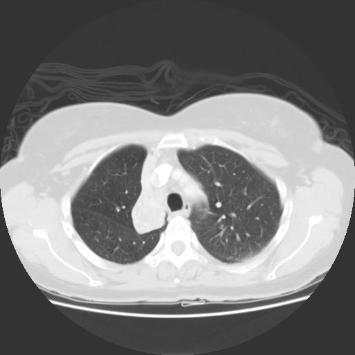
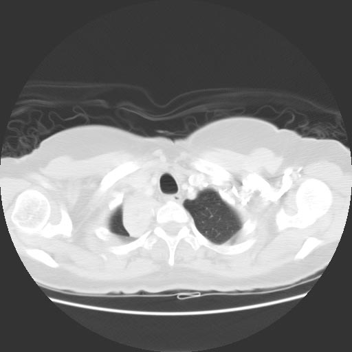
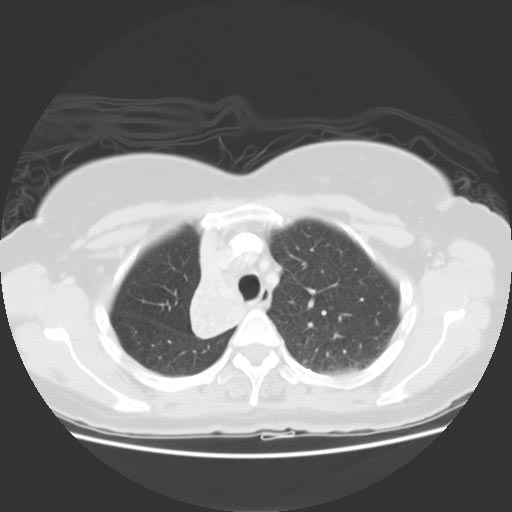
- CT findings associated with right upper lobe (RUL) collapse:
- RUL collapse appears as a right paratracheal opacity
- Minor fissure appears concave laterally
- CT findings associated with atelectatic right middle lobe (RML):
- Tilted icecream sign: Triangular opacity against the border of the right heart with a laterally pointed apex
- CT findings of collapsed right lower lobe (RLL):
- Paraspinal masslike appearance
- CT findings of left upper lobe (LUL) collapse:
- Left upper lobe assumes an inferior location
- Midline shift of RUL
- CT findings of left lower lobe (LLL) collapse:
- Atelectatic LLL is visible in the inferior posterior location
Images shown in this section are courtesy of RadsWiki and copylefted.
-
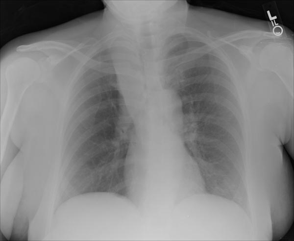
Right upper lobe collapse
-
CT: Right upper lobe collapse


-
CT: Right upper lobe collapse
-

-