Atelectasis CT: Difference between revisions
Ochuko Ajari (talk | contribs) No edit summary |
(→CT) |
||
| (7 intermediate revisions by 2 users not shown) | |||
| Line 1: | Line 1: | ||
__NOTOC__ | __NOTOC__ | ||
{{Atelectasis}} | {{Atelectasis}} | ||
{{CMG}} {{AE}}{{Cherry}} | |||
==Overview== | |||
CT findings suggestive of atelectasis include [[Hilum|hilar]] displacement, elevation of ipsilateral [[Thoracic diaphragm|diaphragm]], rib crowding, [[displacement]] of [[Fissure|fissures]], and compensatory hyperlucency of the remaining [[Lung|lobes]]. CT findings associated with complete atelectasis of an entire [[lung]] include opacification of the entire hemithorax and ipsilateral shift of the [[mediastinum]]. Collapse of different parts of the lung have their own characteristic appearance. For example, a collapsed right middle lobe has a tilted icecream sign which appears as a triangular [[opacity]] against the border of the right heart with a laterally pointed apex. On the other hand, RUL collapse appears as a right paratracheal opacity, with a concave lateral appearance of the [[Fissure|minor lung fissure]]. | |||
{{ | ==CT== | ||
*CT scan may be helpful in the diagnosis of atelectasis. Findings on CT scan suggestive of atelectasis include direct and indirect signs of [[Collapse|lobar collapse]]:<ref name="pmid3279731">{{cite journal |vauthors=Woodring JH |title=Determining the cause of pulmonary atelectasis: a comparison of plain radiography and CT |journal=AJR Am J Roentgenol |volume=150 |issue=4 |pages=757–63 |date=April 1988 |pmid=3279731 |doi=10.2214/ajr.150.4.757 |url=}}</ref> | |||
**Loss of volume on ipsilateral hemithorax | |||
**[[Rib]] crowding | |||
**Silhouetting of the [[Thoracic diaphragm|diaphragm]] or the heart border | |||
**Elevation of ipsilateral [[Thoracic diaphragm|diaphragm]] | |||
**[[Mediastinum|Mediastinal]] shift toward the side of collapse | |||
**[[Hilum|Hilar]] displacement | |||
**Opacification of the [[Collapsed lung|collapsed lobe]] | |||
**[[Displacement]] of [[Fissure|fissures]] | |||
**Compensatory hyperlucency of the remaining [[Lung|lobes]] | |||
*CT findings associated with complete atelectasis of an entire lung:<ref name="pmid4066242">{{cite journal |vauthors=Khoury MB, Godwin JD, Halvorsen RA, Putman CE |title=CT of obstructive lobar collapse |journal=Invest Radiol |volume=20 |issue=7 |pages=708–16 |date=October 1985 |pmid=4066242 |doi= |url=}}</ref> | |||
**Complete collapse of a lung leads to opacification of the entire hemithorax | |||
**Ipsilateral shift of the [[mediastinum]], distinguishing atelectasis from massive [[pleural effusion]] | |||
* CT findings associated with right upper lobe (RUL) collapse:<ref name="pmid6309926">{{cite journal |vauthors=Naidich DP, McCauley DI, Khouri NF, Leitman BS, Hulnick DH, Siegelman SS |title=Computed tomography of lobar collapse: 1. Endobronchial obstruction |journal=J Comput Assist Tomogr |volume=7 |issue=5 |pages=745–57 |date=October 1983 |pmid=6309926 |doi= |url=}}</ref><ref name="pmid8010243">{{cite journal |vauthors=Lee KS, Logan PM, Primack SL, Müller NL |title=Combined lobar atelectasis of the right lung: imaging findings |journal=AJR Am J Roentgenol |volume=163 |issue=1 |pages=43–7 |date=July 1994 |pmid=8010243 |doi=10.2214/ajr.163.1.8010243 |url=}}</ref> | |||
** RUL collapse appears as a right paratracheal opacity | |||
** [[Fissure|Minor fissure]] appears concave laterally | |||
* CT findings associated with atelectatic right middle lobe (RML):<ref name="pmid22282740">{{cite journal |vauthors=Mullett R, Jain A, Kotugodella S, Curtis J |title=Lobar collapse demystified: the chest radiograph with CT correlation |journal=Postgrad Med J |volume=88 |issue=1040 |pages=335–47 |date=June 2012 |pmid=22282740 |doi=10.1136/postgradmedj-2011-130213 |url=}}</ref> | |||
** Tilted icecream sign: Triangular opacity against the border of the right heart with a laterally pointed apex | |||
* CT findings of collapsed right lower lobe (RLL):<ref name="pmid8784731">{{cite journal |vauthors=Molina PL, Hiken JN, Glazer HS |title=Imaging evaluation of obstructive atelectasis |journal=J Thorac Imaging |volume=11 |issue=3 |pages=176–86 |date= 1996 |pmid=8784731 |doi= |url=}}</ref> | |||
** Paraspinal masslike appearance | |||
* CT findings of left upper lobe (LUL) collapse: | |||
** Left upper lobe assumes an inferior location | |||
** Midline shift of RUL | |||
* CT findings of left lower lobe (LLL) collapse: | |||
** Atelectatic LLL is visible in the inferior posterior location | |||
Images shown in this section are courtesy of RadsWiki and copylefted. | Images shown in this section are courtesy of RadsWiki and copylefted. | ||
| Line 29: | Line 59: | ||
[[Category:Pulmonology]] | [[Category:Pulmonology]] | ||
[[Category:Disease]] | [[Category:Disease]] | ||
[[Category:Radiology]] | [[Category:Radiology]] | ||
[[Category:Needs content]] | [[Category:Needs content]] | ||
Latest revision as of 16:09, 22 February 2018
|
Atelectasis Microchapters |
|
Diagnosis |
|---|
|
Treatment |
|
Case Studies |
|
Atelectasis CT On the Web |
|
American Roentgen Ray Society Images of Atelectasis CT |
Editor-In-Chief: C. Michael Gibson, M.S., M.D. [1] Associate Editor(s)-in-Chief: Sudarshana Datta, MD [2]
Overview
CT findings suggestive of atelectasis include hilar displacement, elevation of ipsilateral diaphragm, rib crowding, displacement of fissures, and compensatory hyperlucency of the remaining lobes. CT findings associated with complete atelectasis of an entire lung include opacification of the entire hemithorax and ipsilateral shift of the mediastinum. Collapse of different parts of the lung have their own characteristic appearance. For example, a collapsed right middle lobe has a tilted icecream sign which appears as a triangular opacity against the border of the right heart with a laterally pointed apex. On the other hand, RUL collapse appears as a right paratracheal opacity, with a concave lateral appearance of the minor lung fissure.
CT
- CT scan may be helpful in the diagnosis of atelectasis. Findings on CT scan suggestive of atelectasis include direct and indirect signs of lobar collapse:[1]
- Loss of volume on ipsilateral hemithorax
- Rib crowding
- Silhouetting of the diaphragm or the heart border
- Elevation of ipsilateral diaphragm
- Mediastinal shift toward the side of collapse
- Hilar displacement
- Opacification of the collapsed lobe
- Displacement of fissures
- Compensatory hyperlucency of the remaining lobes
- CT findings associated with complete atelectasis of an entire lung:[2]
- Complete collapse of a lung leads to opacification of the entire hemithorax
- Ipsilateral shift of the mediastinum, distinguishing atelectasis from massive pleural effusion
- CT findings associated with right upper lobe (RUL) collapse:[3][4]
- RUL collapse appears as a right paratracheal opacity
- Minor fissure appears concave laterally
- CT findings associated with atelectatic right middle lobe (RML):[5]
- Tilted icecream sign: Triangular opacity against the border of the right heart with a laterally pointed apex
- CT findings of collapsed right lower lobe (RLL):[6]
- Paraspinal masslike appearance
- CT findings of left upper lobe (LUL) collapse:
- Left upper lobe assumes an inferior location
- Midline shift of RUL
- CT findings of left lower lobe (LLL) collapse:
- Atelectatic LLL is visible in the inferior posterior location
Images shown in this section are courtesy of RadsWiki and copylefted.
-
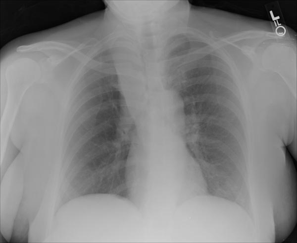
Right upper lobe collapse
-
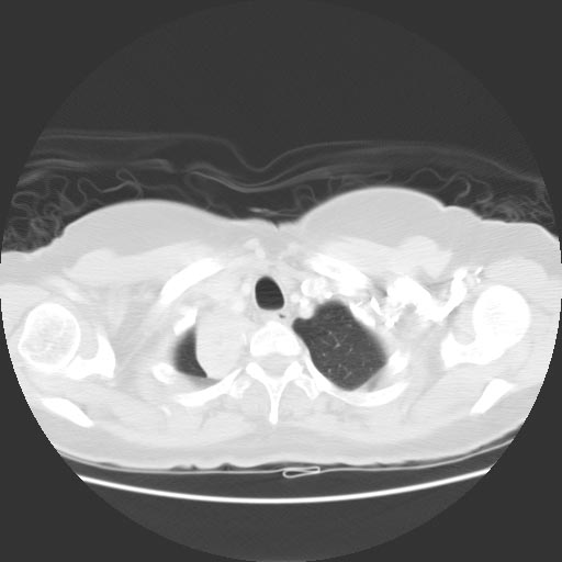
CT: Right upper lobe collapse


-
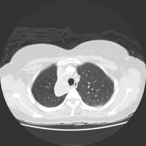
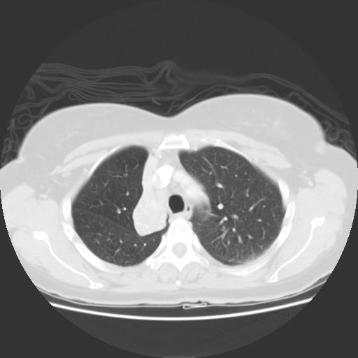
CT: Right upper lobe collapse
-

-


References
- ↑ Woodring JH (April 1988). "Determining the cause of pulmonary atelectasis: a comparison of plain radiography and CT". AJR Am J Roentgenol. 150 (4): 757–63. doi:10.2214/ajr.150.4.757. PMID 3279731.
- ↑ Khoury MB, Godwin JD, Halvorsen RA, Putman CE (October 1985). "CT of obstructive lobar collapse". Invest Radiol. 20 (7): 708–16. PMID 4066242.
- ↑ Naidich DP, McCauley DI, Khouri NF, Leitman BS, Hulnick DH, Siegelman SS (October 1983). "Computed tomography of lobar collapse: 1. Endobronchial obstruction". J Comput Assist Tomogr. 7 (5): 745–57. PMID 6309926.
- ↑ Lee KS, Logan PM, Primack SL, Müller NL (July 1994). "Combined lobar atelectasis of the right lung: imaging findings". AJR Am J Roentgenol. 163 (1): 43–7. doi:10.2214/ajr.163.1.8010243. PMID 8010243.
- ↑ Mullett R, Jain A, Kotugodella S, Curtis J (June 2012). "Lobar collapse demystified: the chest radiograph with CT correlation". Postgrad Med J. 88 (1040): 335–47. doi:10.1136/postgradmedj-2011-130213. PMID 22282740.
- ↑ Molina PL, Hiken JN, Glazer HS (1996). "Imaging evaluation of obstructive atelectasis". J Thorac Imaging. 11 (3): 176–86. PMID 8784731.