Anaplastic thyroid cancer pathophysiology
|
Anaplastic thyroid cancer Microchapters |
|
Differentiating Anaplastic thyroid cancer from other Diseases |
|---|
|
Diagnosis |
|
Treatment |
|
Case Studies |
|
Anaplastic thyroid cancer pathophysiology On the Web |
|
American Roentgen Ray Society Images of Anaplastic thyroid cancer pathophysiology |
|
Risk calculators and risk factors for Anaplastic thyroid cancer pathophysiology |
Editor-In-Chief: C. Michael Gibson, M.S., M.D. [1]; Associate Editor(s)-in-Chief: Ammu Susheela, M.D. [2]
Overview
Anaplastic thyroid cancer arises from cells of the thyroid, which are normally involved in production and secretion of thyroid hormones, thyroxine (T4) and triiodothyronine (T3). Anaplastic thyroid tumor is always considered as stage IV. Genes involved in the pathogenesis of follicular thyroid cancer include P53 and BRAF.
Pathogenesis
- Anaplastic tumors have a high mitotic rate and lymphovascular invasion. It rapidly invades surrounding tissues (such as the trachea). The presence of regional lymphadenopathy in older patients in whom needle aspiration biopsy reveals characteristic vesicular appearance of the nuclei would support a diagnosis of anaplastic carcinoma. Most cases of anaplastic thyroid cancer have distant metastasis at the time of diagnosis because of the rapid growth and aggressive nature of this type of cancer. Common sites of metastasis include the lungs, pleura, bones, and brain.
- It is always considered as stage IV.
Genetics
- Mutation of P53 tumor suppressor gene is associated with anaplastic thyroid cancer.[1]
- Mutation of BRAF gene is also associated with anaplastic thyroid cancer.
Associated Conditions
Gross Pathology
- Unencapsulated, fleshy, tan-white tumor
- Soft-tissue infiltration of the neck
Microscopic Pathology
- Three types of histologic variety
- Spindle
- Giant cell
- Squamoid cell
Features:
- Cytologically malignant:
- Huge nuclear-cytoplasmic ratio
- Mitoses
- Presence or absence of necrosis
-
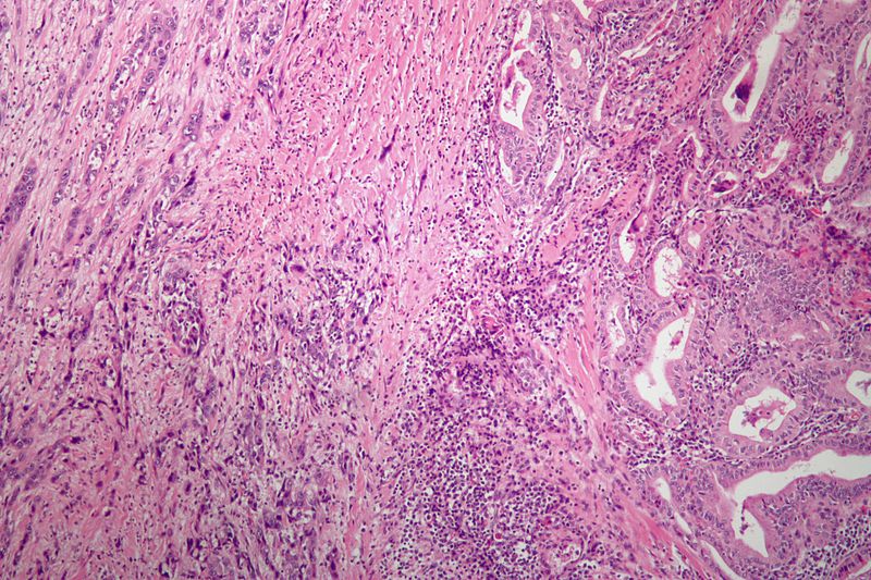
Anaplastic thyroid carcinoma with a component of PTC. (WC)

References
- ↑ Patel KN, Shaha AR (2006). "Poorly differentiated and anaplastic thyroid cancer". Cancer Control. 13 (2): 119–28. PMID 16735986.