Amoebiasis pathophysiology
|
Amoebiasis Microchapters |
|
Diagnosis |
|---|
|
Treatment |
|
Case Studies |
|
Amoebiasis pathophysiology On the Web |
|
American Roentgen Ray Society Images of Amoebiasis pathophysiology |
|
Risk calculators and risk factors for Amoebiasis pathophysiology |
Editor-In-Chief: C. Michael Gibson, M.S., M.D. [1]; Associate Editor(s)-in-Chief: Jesus Rosario Hernandez, M.D. [2]
Overview
Symptoms can range from mild diarrhoea to dysentery with blood and mucus. The blood comes from amoebae invading the lining of the intestine. In about 10% of invasive cases the amoebae enter the bloodstream and may travel to other organs in the body. Most commonly this means the liver, as this is where blood from the intestine reaches first, but they can end up almost anywhere.
Pathophysiology
In asymptomatic infections the amoeba lives by eating and digesting bacteria and food particles in the gut. It does not usually come in contact with the intestine itself due to the protective layer of mucus that lines the gut. Disease occurs when amoeba comes in contact with the cells lining the intestine. It then secretes the same substances it uses to digest bacteria, which include enzymes that destroy cell membranes and proteins. This process leads to penetration and digestion of human tissues, resulting first in flask-shaped ulcers in the intestine. Entamoeba histolytica ingests the destroyed cells by phagocytosis and is often seen with red blood cells inside. Especially in Latin America, a granulomatous mass (known as an amoeboma) may form in the wall of the colon due to long-lasting cellular response, and is sometimes confused with cancer.
Theoretically, the ingestion of one viable cyst can cause an infection.
Colon: Amebiasis
{{#ev:youtube|Xti6OURHxhc}}
Transmission
Amoebiasis is usually transmitted by the fecal-oral route, but it can also be transmitted indirectly through contact with dirty hands or objects as well as by oral-anal contact.
Amoebiasis is usually transmitted by the fecal-oral route (contamination of drinking water and foods with fecal matter), but it can also be transmitted indirectly through contact with dirty hands or objects as well as by anal-oral contact.
Infection is spread through ingestion of the cyst form of the parasite, a semi-dormant and hardy structure found in feces. Free-living amoebae, or trophozoites, that do not form cysts but die quickly after leaving the body may also be present: these are rarely the source of new infections.
Amoebic dysentery is often confused with "traveler's diarrhea", or "Montezuma's Revenge" in Mexico, because of the prevalence of both in developing nations. In fact, most traveler's diarrhea is bacterial or viral in origin.
Liver abscesses can occur without previous development of amoebic dysentery.
Life Cycle
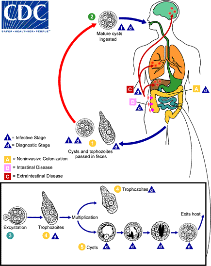
Cysts and trophozoites are passed in feces (1). Cysts are typically found in formed stool, whereas trophozoites are typically found in diarrheal stool. Infection by Entamoeba histolytica occurs by ingestion of mature cysts (2) in fecally contaminated food, water, or hands. Excystation (3) occurs in the small intestine and trophozoites (4) are released, which migrate to the large intestine. The trophozoites multiply by binary fission and produce cysts (5), and both stages are passed in the feces (1). Because of the protection conferred by their walls, the cysts can survive days to weeks in the external environment and are responsible for transmission. Trophozoites passed in the stool are rapidly destroyed once outside the body, and if ingested would not survive exposure to the gastric environment. In many cases, the trophozoites remain confined to the intestinal lumen (A: noninvasive infection) of individuals who are asymptomatic carriers, passing cysts in their stool. In some patients the trophozoites invade the intestinal mucosa (B: intestinal disease), or, through the bloodstream, extraintestinal sites such as the liver, brain, and lungs (C: extraintestinal disease), with resultant pathologic manifestations. It has been established that the invasive and noninvasive forms represent two separate species, respectively E. histolytica and E. dispar. These two species are morphologically indistinguishable unless E. histolytica is observed with ingested red blood cells (erythrophagocystosis). Transmission can also occur through exposure to fecal matter during sexual contact (in which case not only cysts, but also trophozoites could prove infective).
-
Life cycle of Amebiasis
Adapted from CDC
Gallery
-
Invasive extraintestinal amebiasis. Adapted from Public Health Image Library (PHIL). [1]
-
Amebic abscess of liver. Adapted from Public Health Image Library (PHIL). [1]
-
Intestinal ulcers due to amebiasis. Adapted from Public Health Image Library (PHIL). [1]
-
Intestinal ulcers due to amebiasis. Adapted from Public Health Image Library (PHIL). [1]
-
Intestinal amebiasis. Adapted from Public Health Image Library (PHIL). [1]
-
Amebic abscess in liver. Adapted from Public Health Image Library (PHIL). [1]
-
Amebic abscess in liver. Adapted from Public Health Image Library (PHIL). [1]
-
Amebiasis in intestine. Adapted from Public Health Image Library (PHIL). [1]
References

![Invasive extraintestinal amebiasis. Adapted from Public Health Image Library (PHIL). [1]](/images/4/48/Amebiasis04.jpeg)
![Amebic abscess of liver. Adapted from Public Health Image Library (PHIL). [1]](/images/e/ef/Amebiasis11.jpeg)
![Intestinal ulcers due to amebiasis. Adapted from Public Health Image Library (PHIL). [1]](/images/b/b0/Amebiasis12.jpeg)
![Intestinal ulcers due to amebiasis. Adapted from Public Health Image Library (PHIL). [1]](/images/b/b0/Amebiasis13.jpeg)
![Intestinal amebiasis. Adapted from Public Health Image Library (PHIL). [1]](/images/7/72/Amebiasis15.jpeg)
![Amebic abscess in liver. Adapted from Public Health Image Library (PHIL). [1]](/images/c/cf/Amebiasis16.jpeg)
![Amebic abscess in liver. Adapted from Public Health Image Library (PHIL). [1]](/images/b/b6/Amebiasis17.jpeg)
![Amebiasis in intestine. Adapted from Public Health Image Library (PHIL). [1]](/images/1/1c/Amebiasis18.jpeg)