Scleroderma physical examination
|
Scleroderma Microchapters |
|
Diagnosis |
|---|
|
Treatment |
|
Case Studies |
|
Scleroderma physical examination On the Web |
|
American Roentgen Ray Society Images of Scleroderma physical examination |
|
Risk calculators and risk factors for Scleroderma physical examination |
Editor-In-Chief: C. Michael Gibson, M.S., M.D. [1]
Overview
Physical Examination
Skin
-
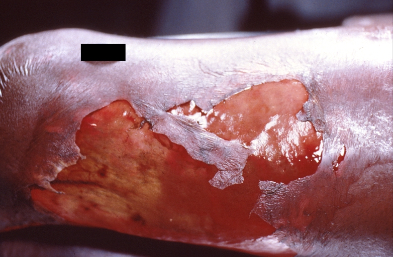
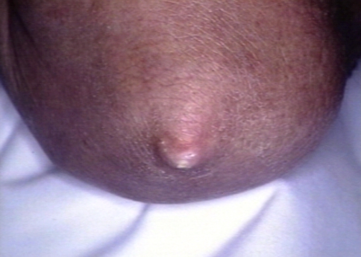
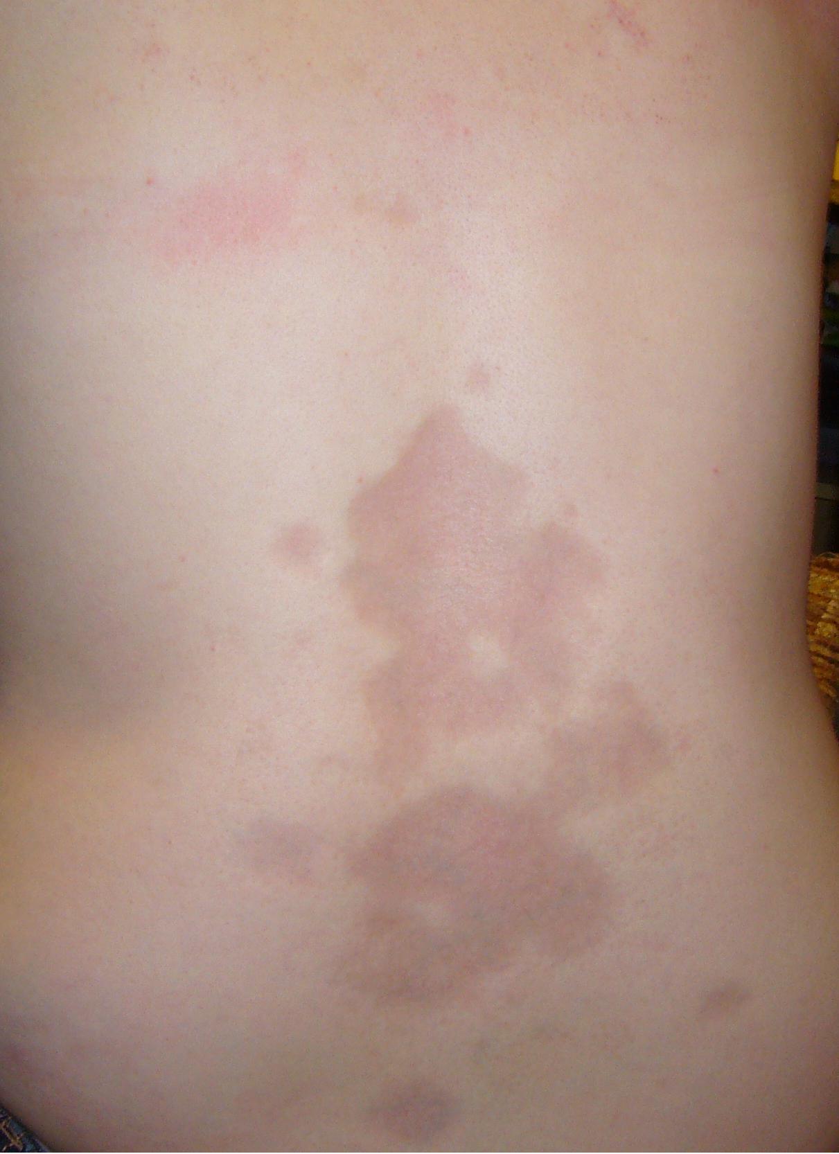
Panniculitis and fascitis, streptococcus A septicemia in a patient with Scleroderma who was on high dose steroids
-
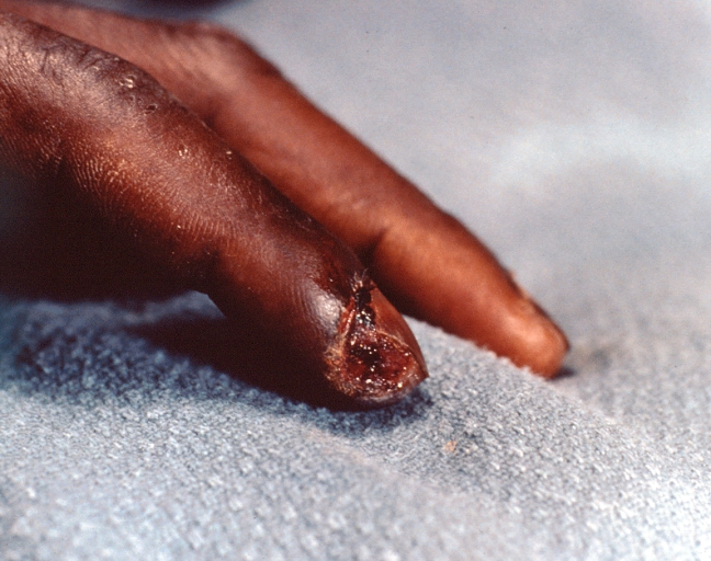
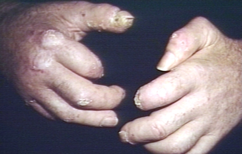
Necrosis of distal finger in a patient with panniculitis and fascitis, streptococcus A septicemia in a patient with Scleroderma who was on high dose steroids
-
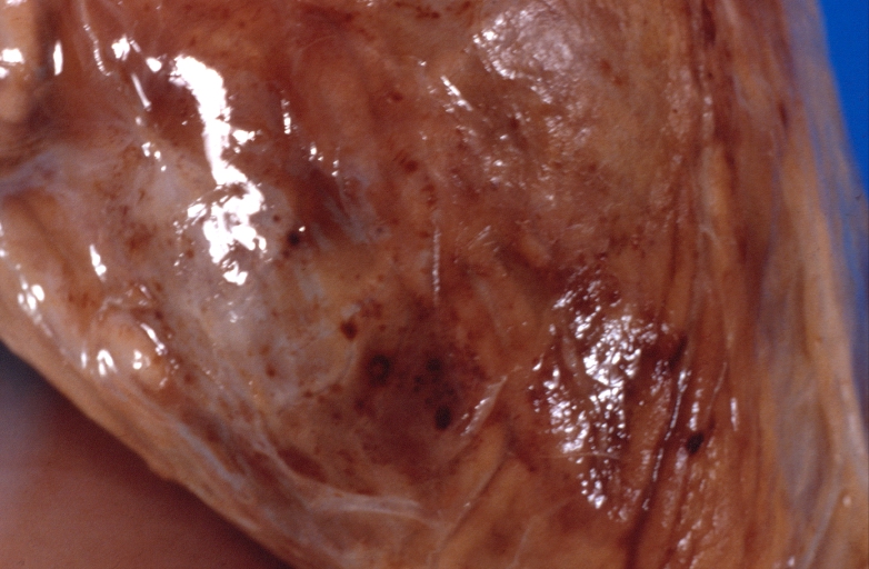
Panniculitis and fascitis, streptococcus A septicemia in a patient with Scleroderma who was on high dose steroids
-
Skin: Scleroderma; shoulders & back
-
Skin: Scleroderma in crest syndrome; calcinosis at elbow
-
Hands: Scleroderma,
-
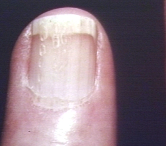
Hand: Scleroderma, finger, posterior nail fold
-
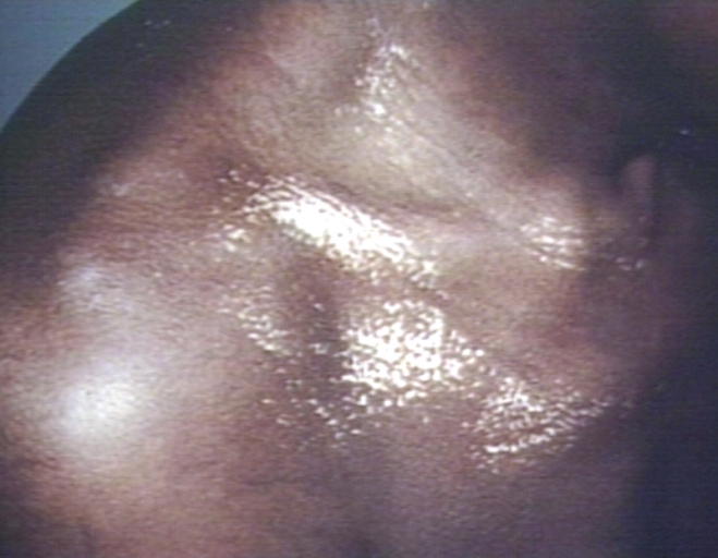
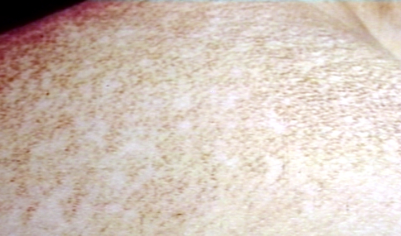
Skin: Scleroderma, chest, salt and pepper pigmentation
-
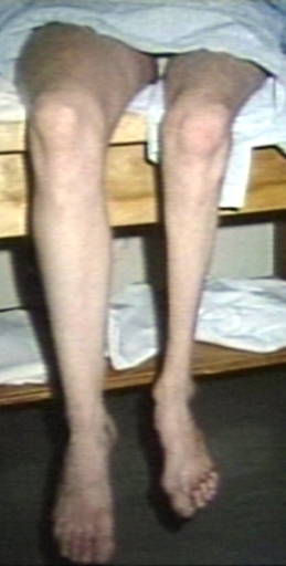
Leg: Morphea, circumscribed scleroderma; age 19
-
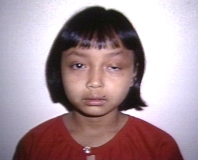
Face: Morphea, circumscribed scleroderma
-
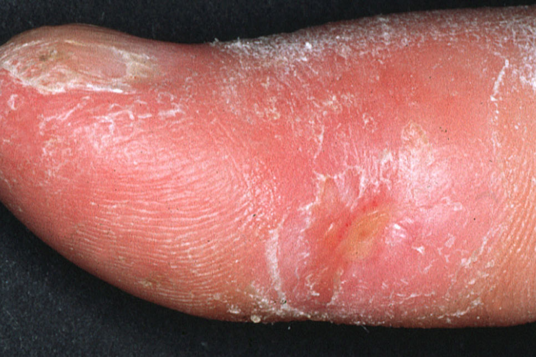
Clinical appearance of acrosclerotic piece-meal necrosis of the thumb in a patient with systemic sclerosis.
-
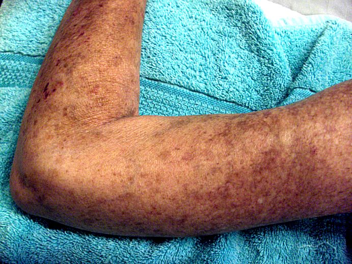
Left arm of Scleroderma patient, showing skin lesions
-
Morphea


Sources
Image courtesy of Professor Peter Anderson DVM PhD and published with permission © PEIR, University of Alabama at Birmingham, Department of Pathology [2]