Renal artery stenosis pathophysiology
|
Renal artery stenosis Microchapters |
|
Diagnosis |
|---|
|
Treatment |
|
Case Studies |
|
Renal artery stenosis pathophysiology On the Web |
|
American Roentgen Ray Society Images of Renal artery stenosis pathophysiology |
|
Risk calculators and risk factors for Renal artery stenosis pathophysiology |
Editor-In-Chief: C. Michael Gibson, M.S., M.D. [1] Associate Editor(s)-in-Chief: Shivam Singla, M.D.[2]
Overview
The reduction in renal blood flow secondary to renal artery stenosis stimulates renin release from the juxtaglomerular apparatus through activation of the tubuloglomerular feedback, baroreceptor reflex, and the sympathetic nervous system. Elevated angiotensin II activities in turn cause elevation of the arterial pressure and other effects including aldosterone secretion, sodium retention, and left ventricular hypertrophy and remodeling.[1]
Pathophysiology
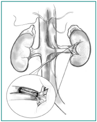
Renal artery stenosis means the narrowing of both renal arteries leading to the on=bstruction of blood flow and resulting in the release of renin and activation of the renin-angiotensin-aldosterone system. This leads to the increase in production of aldosterone promoting increased retention of sodium and an increase in peripheral vascular resistance leading to increase renovascular hypertension that is also called secondary hypertension. Prolonged hypoperfusion to the kidneys resulting in chronic stimulation and hyperplasia of the juxtaglomerular apparatus. This prolonged ischemia further leads to renal insufficiency and inturn progressive renal atrophy. In atherosclerosis, the initiator of endothelial injury although not well understood can be, dyslipidemia, cigarette smoking, viral infection, immune injury, or increased homocysteine levels. [5]At the lesion site, permeability to low-density lipoprotein (LDL) and macrophage migration increases with subsequent proliferation of endothelial and smooth muscle cells and ultimate formation of atherosclerotic plaque. Renal blood flow, which is significantly greater than the perfusion to other organs, along with glomerular capillary hydrostatic pressure is an important determinant of the glomerular filtration rate (GFR). In patients with renal artery stenosis, the chronic ischemia produced by the obstruction of renal blood flow leads to adaptive changes in the kidney which include the formation of collateral blood vessels and secretion of renin by juxtaglomerular apparatus. The renin enzyme has an important role in maintaining homeostasis in that it converts angiotensinogen to angiotensin I. Angiotensin I has then converted to angiotensin II with the help of an angiotensin-converting enzyme (ACE) in the lungs. Angiotensin II is responsible for vasoconstriction and release of aldosterone which causes sodium and water retention, thus resulting in secondary hypertension or renovascular hypertension.[1][6][1]
Pathogenesis of Chronic Renal Insufficiency
Glomerular filtration rate (GFR) is autoregulated by angiotensin II and other modulators between the afferent and efferent arteries. The maintenance of GFR fails when renal perfusion pressure falls below 70 mmHg to -85 mmHg. Therefore significant functional impairment of autoregulation, leading to a decrease in the GFR, is only likely to be observed until arterial luminal narrowing exceeds 50%. Studies demonstrate that a moderate reduction in renal perfusion pressure (up to 40%) and renal blood flow (mean 30%) cause reduction in glomerular filtration, however, tissue oxygenation within the kidney cortex and medulla can adapt without the development of severe hypoxia. As an inference patients at an early stage can be treated with medical therapy without progressive loss of function or irreversible fibrosis in many cases, sometimes for many years.
It is reported that more advanced stenosis corresponding to a 70% to 80% of vascular occlusion leads to demonstrable cortical hypoxia, and it is proposed that this hypoxia produce rarefaction of microvessels, as well as activation of inflammatory and oxidative pathways that cause interstitial fibrosis. [7]Therefore loss of renal function in renovascular disease in addition to being a usually reversible consequence of antihypertensive therapy can reflect a progressive narrowing of the renal arteries and/or progressive intrinsic renal disease. Eventually, long-standing parenchymal injury becomes an irreversible process. At this point, restoring renal blood flow provides no recovery of renal function or clinical benefit.
