Sandbox 2
Lower GI bleeding is defined as any bleed that occurs distal to the ligament of Treitz.
Incidence
- In the United States the incidence of LGIB ranges from 20.5 to 27 per 100,000 persons per year.
Age
- There is a greater than 200 fold increase from the third to the ninth decade of life.
Classification
- Lower GI bleeding can be classified into 3 groups based on the severity of bleeding:
- Occult lower GI bleeding
- Moderate lower GI bleeding
- Severe lower GI bleeding
Occult lower GI bleeding
- Occult lower GI bleeds can present in patients at any age.
- Lab work reveals patients with microcytic hypochromic anemia due to chronic blood loss.
- The differential diagnosis of these patients should include inflammatory, neoplastic and congenital.
- The patient typically appears well, hemodynamically stable.
Moderate lower GI bleeding
- Moderate bleeding can occur at any age and presents as hematochezia or melena.
- The patient is usually hemodynamically stable.
- Many disease processes should be considered on the differential list including neoplastic disease, inflammatory, infectious, benign anorectal, and congenital.
Severe lower GI bleeding
- Massive bleeding usually occurs in patients older than 65 years with multiple medical problems, and this bleeding presents as hematochezia or bright red blood per rectum.
- The patient is usually hemodynamically unstable with a systolic blood pressure (SBP) equal to or less than 90 mmHg, heart rate (HR) less than or equal 100/min, and low urine output.
- Lab work reveals a hemoglobin equal to or less than 6 g/dl.
- Massive lower GI bleeds are mostly due to diverticulosis and angiodysplasias. The mortality rate may be as high as 21%.
| Severe lower GI bleeding | Moderate lower GI bleeding | Occult lower GI bleeding | |
|---|---|---|---|
| Age | > 65 years | occur at any age | any age. |
| Presenting symptoms | Hematochezia or bright red blood per rectum. | Hematochezia or melena. | |
| Hemodynamics | Unstable | Stable | Stable |
| Lab findings | hemoglobin equal to or less than 6 g/dl. | Microcytic anemia | Microcytic hypochromic anemia due to chronic blood loss. |
| Differential | Diverticulosis and angiodysplasias | Neoplastic disease Inflammatory, <br> infectious, benign anorectal, and congenital diseases. | Inflammatory, neoplastic and congenital. |
Blood supply
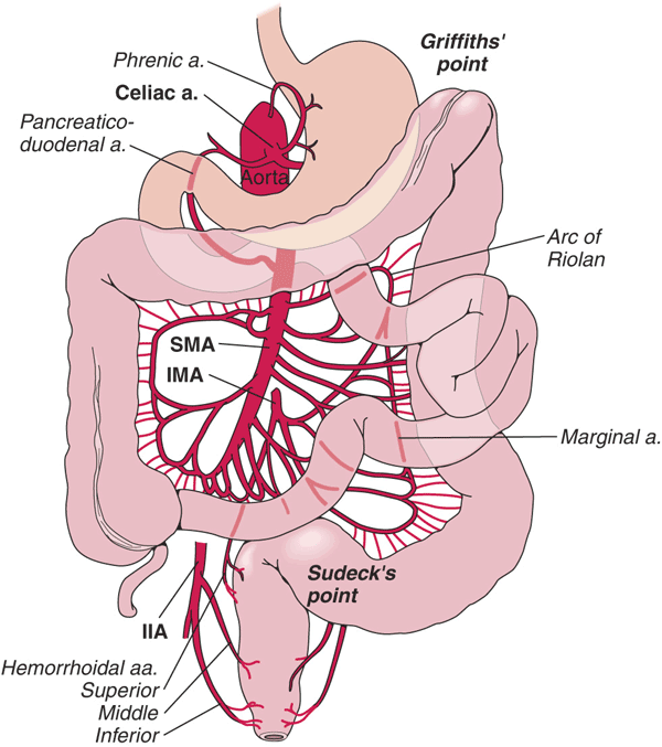
- The SMA and IMA are connected by the marginal artery of Drummond.
- This vascular arcade runs in the mesentery close to the bowel.
- As patients age, there is increased incidence of occlusion of the IMA.
- The left colon stays perfused, primarily because of the marginal artery.
| Lower GI Tract | Arterial Supply | Venous Drainage | |
|---|---|---|---|
| Midgut |
|
|
|
| Hindgut |
|
|
|
| ɸ -Except lower rectum, which drains into the systemic circulation. | |||

Source: By Anpol42 (Own work) [CC BY-SA 4.0 (https://creativecommons.org/licenses/by-sa/4.0)], via Wikimedia Commons
Pathogenesis
Diverticulosis is the most common etiology of lower GI bleeding accounting for 30% of all cases, followed by anorectal disease, ischemia, inflammatory bowel disease (IBD), neoplasia and arteriovenous (AV) malformations.
- Diverticulosis
- The colonic wall weakens with age and results in the formation of saclike protrusions known as diverticula.
- These protrusions generally occur at the junction of blood vessel penetrating through the mucosa and circular muscle fibers of the colon.
- Diverticula are most common in the descending and sigmoid colon.
- Despite the majority of diverticula being on the left side of the colon, diverticular bleeding originates from the right side of the colon in 50% to 90% of instances.
- Most of the time bleeding from diverticulosis stops spontaneously, however, in about 5% of patients, the bleeding can be massive and life-threatening.

Source:By Anpol42 (Own work) [CC BY-SA 4.0 (https://creativecommons.org/licenses/by-sa/4.0)], via Wikimedia Commons
- Anorectal disease
- Hemorrhoids and anal fissures are the most common disease under anorectal disease responsible for GI bleeding.
- Hemorrhoids are engorged vessels in the normal anal cushions. When swollen, this tissue is very friable and susceptible to trauma, which leads to painless, bright red bleeding.
- Anal fissures are defined as a tear in the anal mucosa. With the passage of stool, the mucosa continues to tear and leads to bright red bleeding.
- Mesenteric Ischemia
- Mesenteric ischemia results when there is inadequate blood supply at the level of the small intestine.
- 2 or more vessels (celiac, SMA, or IMA) must be involved for symptoms to occur.
- Non Occlusive MI affects critically ill patients who are vasopressor-dependent.
- Venous thrombosis of the visceral vessels can also precipitate an acute ischemic event.
- Ischemic Colitis
- Ischemic colitis is caused by poor perfusion of the colon, which results in the inability of that area of the colon to meet its metabolic demands.
- It can be gangrenous or nongangrenous, acute, transient, or chronic.
- The left colon is predominantly affected, with the splenic flexure having increased susceptibility.
- Intraluminal hemorrhage occurs as the mucosa becomes necrotic, sloughs, and bleeds.
- Damage to the tissue is caused both with the ischemic insult as well as reperfusion injury.
- Inflammatory Bowel Disease
- In Crohn's disease T cell activation stimulates interleukin (IL)-12 and tumor necrosis factor (TNF)-a, which causes chronic inflammation and tissue injury.
- Initially, inflammation starts focally around the crypts, followed by superficial ulceration of the mucosa.
- The deep mucosal layers are then invaded in a noncontinuous fashion, and noncaseating granulomas form, which can invade through the entire thickness of the bowel and into the mesentery and surrounding structures.
- In ulcerative colitis T cells cytotoxic to the colonic epithelium accumulate in the lamina propria, accompanied by B cells that secrete immunoglobulin G (IgG) and IgE.
- This results in inflammation of the crypts of Lieberkuhn, with abscesses and pseudopolyps.
- Ulcerative colitis generally begins at the rectum and is a continuous process confined exclusively to the colon.
- Neoplasia
- Colon carcinoma follows a distinct progression from polyp to cancer.
- Mutations of multiple genes are required for the formation of adenocarcinoma, including the APC gene, Kras, DCC, and p53.
- Certain hereditary syndromes are also classified by defects in DNA mismatch repair genes and microsatellite instability.
- These tumors tend to bleed slowly, and patients present with hemocult positive stools and microcytic anemia.
- Although cancers of the small bowel are much less common than colorectal cancers, they should be ruled out in cases of lower GI bleeding in which no other source is identified.
- AV Malformation/Angiodysplasia
- In AV malformation direct connections between arteries and veins occur in the colonic submucosa.
- The lack of capillary buffers causes high pressure blood to enter directly into the venous system, making these vessels at high risk of rupture into the bowel lumen.
- In Angiodysplasia over time, previously healthy blood vessels of the cecum and ascending colon degenerate and become prone to bleeding.
- Although 75% of angiodysplasia cases involve the right colon, they are a significant cause of obscure bleeding and the most common cause of bleeding from the small bowel in the elderly.
Epidemiology
Prevalence
- Approximately 20 patients/100,000 population in the U.S.
Incidence
- The estimated annual incidence of lower GI bleeding is approximately 0.03% in the adult population as a whole.
Demographics
Gender
- More common in men than women
Age
- Rare in children
- The incidence of lower GI bleeding increases with age with a 200-fold increase from the second to eighth decades of life la.
- Largely due to the increase in the prevalence of diverticular disease and angiodysplasia with age.
Symptoms
- Occult LGIB may present with symptoms of iron deficiency anemia such as fatigue, palpitations, and dyspnea.
- Patients with intussusception may present with pallor and vomiting in addition to LGIB
- Associated symptoms, such as abdominal pain or change in bowel habits, may also aide in the diagnosis
- Bloody diarrhea associated with abdominal pain may suggest an infectious cause or IBD in a younger patient and ischemic colitis in an older patient with vascular disease
- Painless bleeding usually suggests angiodysplasia, diverticular disease, or internal hemorrhoids
- Perianal pain suggests a perianal fissure or fistula
History
- A detailed description of the nature of the blood loss can help ascertain the likely source of bleeding
- The clinical history should identify whether this is a recurrent bleed.
- Bleeding from angiodysplasia is usually recurrent and chronic, but severe bleeding resulting in hemodynamic instability can occur
- Associated weight loss suggests malignancy
- A history of recent colonic polypectomy or biopsy indicates iatrogenic bleeding.
- This is usually low grade and limited, although it can be severe if an underlying artery is involved or if there is an inadequate coagulation of the polypectomy stalk.
- In 1.5% of polypectomies bleeding occurs immediately. However, delayed bleeding can occur several hours or days following the procedure
- It is essential to establish the presence of comorbid diseases, as these not only help in diagnosis but may also influence treatment.
- The presence of systemic diseases such as atherosclerotic disease, IBD, coagulopathies, and HIV, and a history of pelvic irradiation for malignancy should be considered
- A family history of disease such as IBD or colorectal malignancy is relevant