Unused files
Jump to navigation
Jump to search
The following files exist but are not embedded in any page. Please note that other web sites may link to a file with a direct URL, and so may still be listed here despite being in active use.
Showing below up to 50 results in range #251 to #300.
-
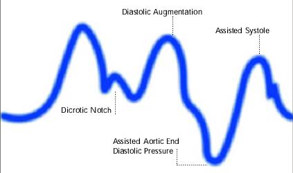
 Late deflation.JPG 423 × 259; 12 KB
-
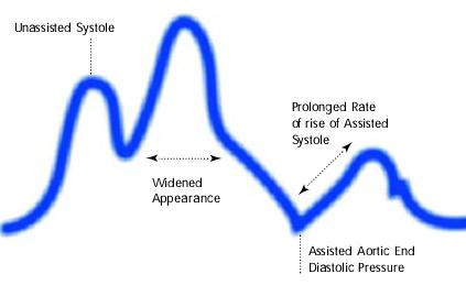
 Late inflation.JPG 423 × 250; 12 KB
-
 MOV00019.swf ; 1.18 MB
-
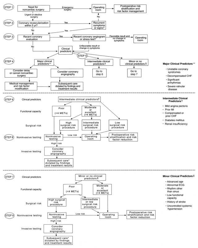 Preoperative Clearance.jpg 640 × 799; 60 KB
-
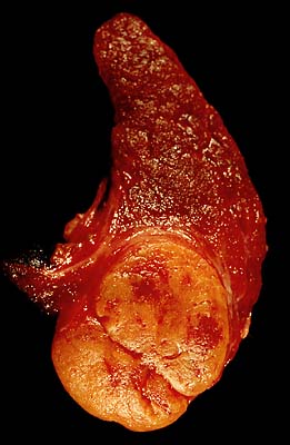 Follicular adenoma of the thyroid.jpg 261 × 400; 23 KB
-
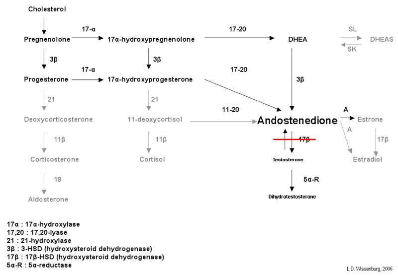 800px-Test biosynth 17BHSD3.jpg 800 × 556; 38 KB
-
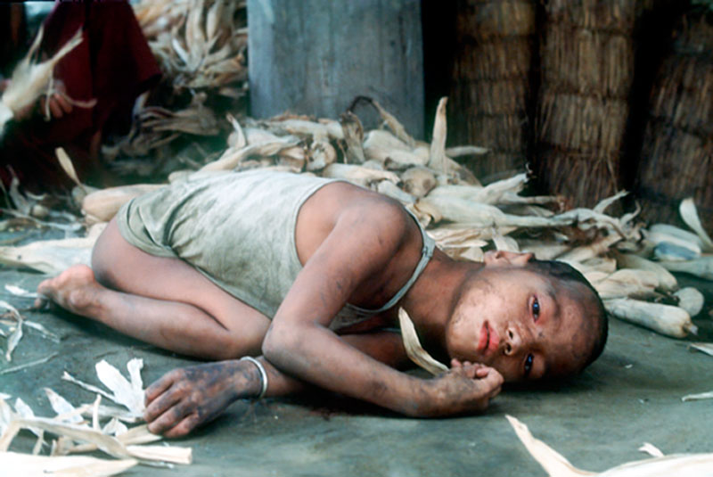 Cretin Child (1).jpg 800 × 535; 108 KB
-
 560px-DHEA1.svg.png 560 × 225; 16 KB
-
 DHEA1.svg.png 560 × 225; 16 KB
-
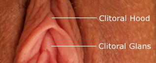 Hood and glans labeled.jpg 317 × 129; 34 KB
-
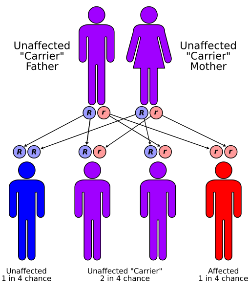 Autorecessive.svg.png 512 × 599; 52 KB
-
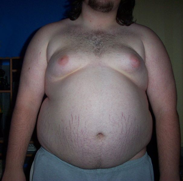 Central Obesity 008.jpg 606 × 600; 38 KB
-
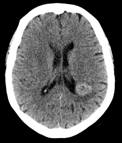 Brain met.jpg 511 × 599; 39 KB
-
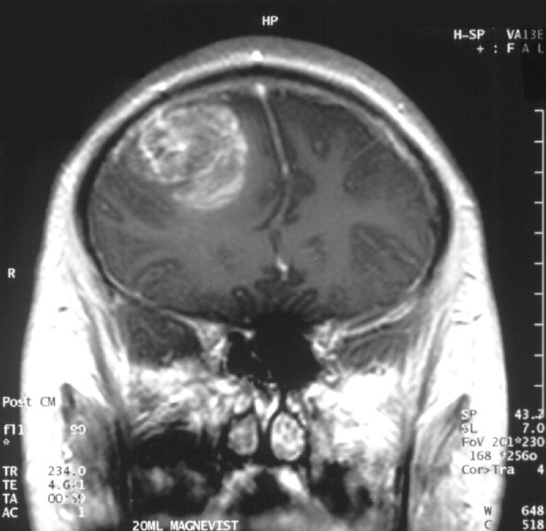 Glioblastoma - MR coronal with contrast.jpg 617 × 600; 57 KB
-
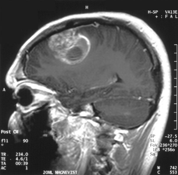 Glioblastoma - MR sagittal with contrast.jpg 612 × 599; 59 KB
-
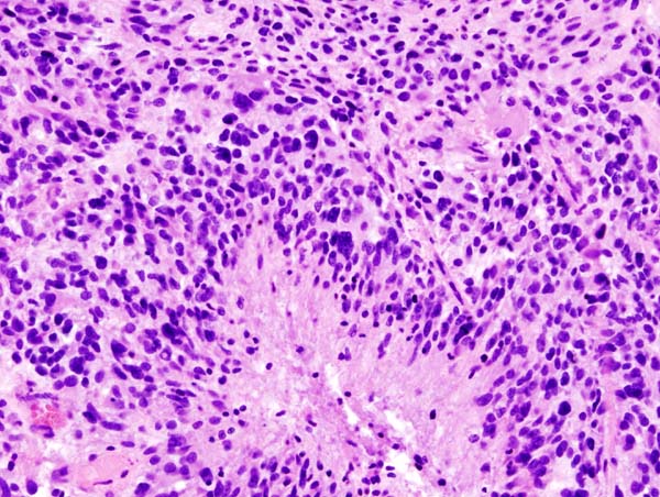 Glioblastoma (1).jpg 600 × 452; 94 KB
-
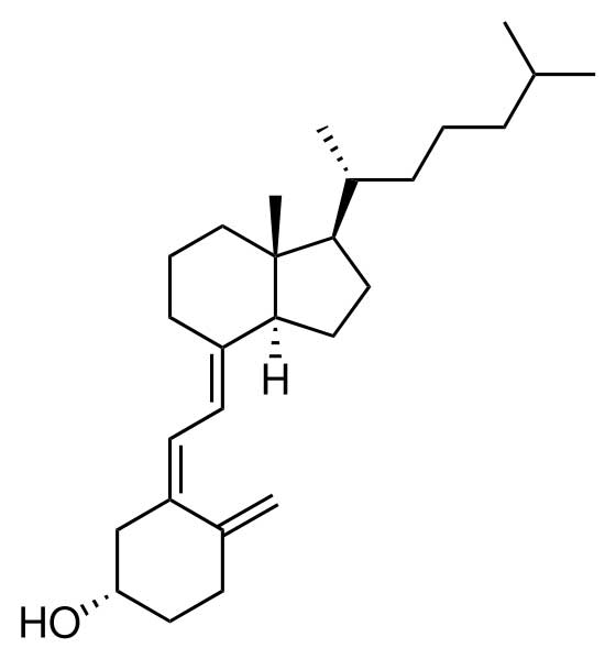 Cholecalciferol.jpg 560 × 599; 10 KB
-
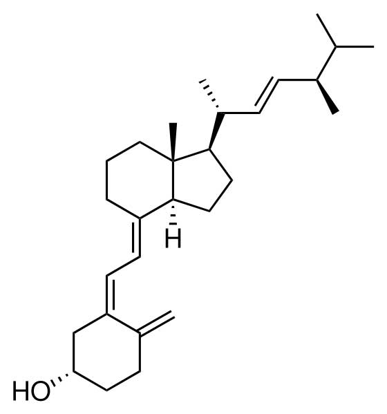 Ergocalciferol.svg.jpg 560 × 599; 11 KB
-
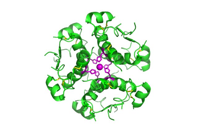 InsulinHexamer.jpg 640 × 434; 41 KB
-
 BCtimeline.jpg 800 × 481; 71 KB
-
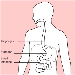 Stomach diagram.svg.png 255 × 255; 22 KB
-
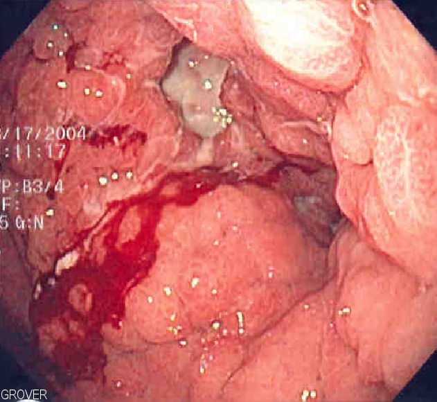 Linitis plastica 2.jpg 631 × 580; 77 KB
-
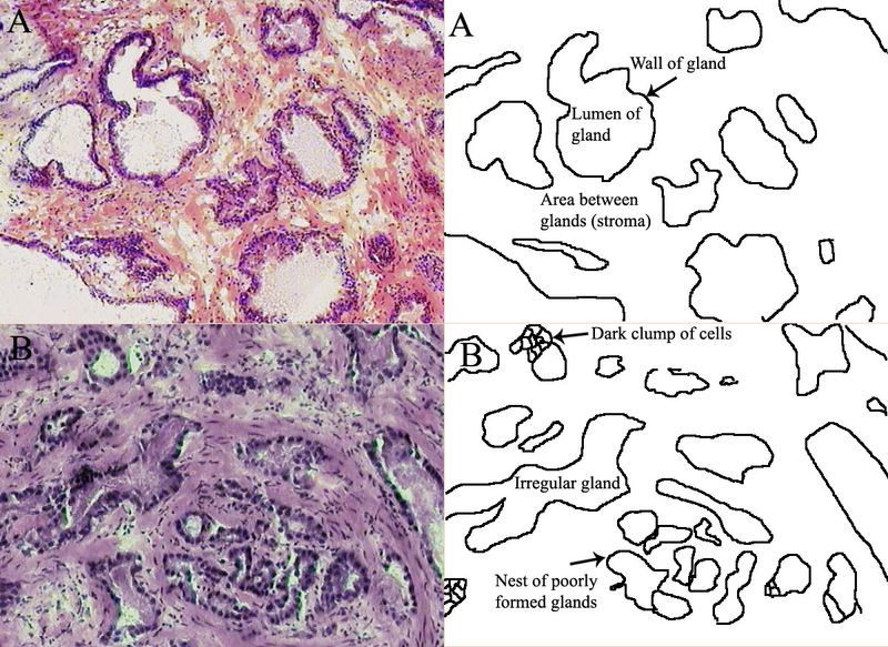 Prostatehistopath.jpg 800 × 583; 165 KB
-
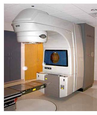 Linacprostate.jpg 324 × 385; 19 KB
-
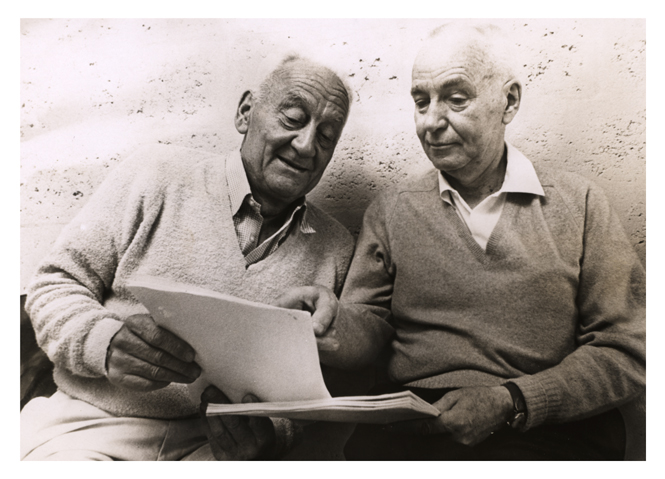 Hugginsprostate.jpg 665 × 479; 217 KB
-
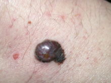
 Malignant melanoma.jpg 320 × 240; 154 KB
-
 WB032021.JPG 800 × 532; 73 KB
-
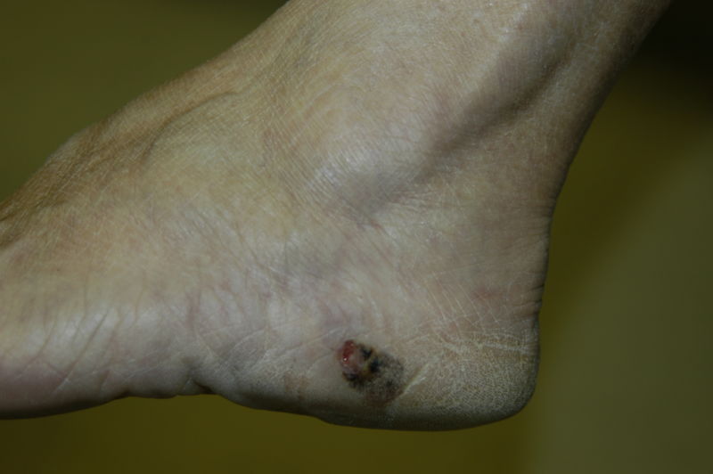
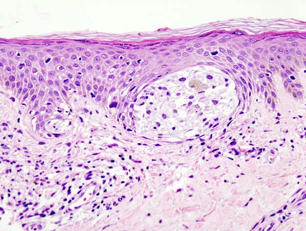 Malignant melanoma (1) at thigh Case 01.jpg 600 × 452; 72 KB
-
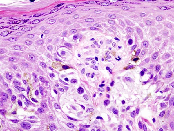 Malignant melanoma (4) at thigh Case 01.jpg 600 × 452; 72 KB
-
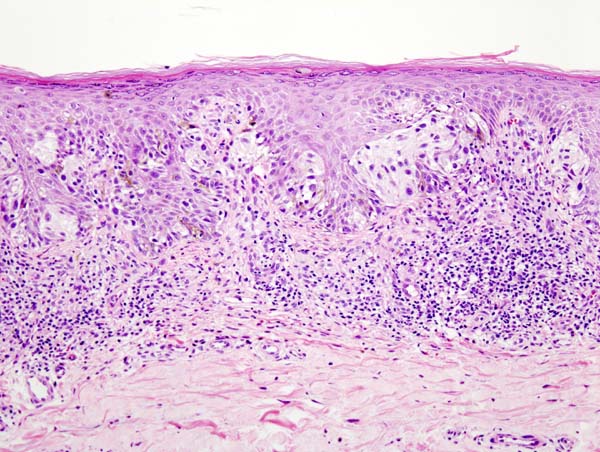
 Superficial spreading melanoma 1 060619.jpg 220 × 165; 41 KB
-
 MMatthigh.jpg 600 × 452; 84 KB
-
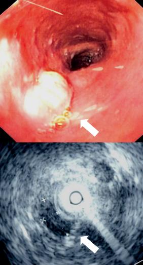 Mid esophageal mass.jpg 282 × 523; 24 KB
-
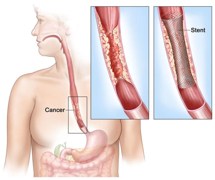 Esophagael stent.jpg 715 × 599; 61 KB
-
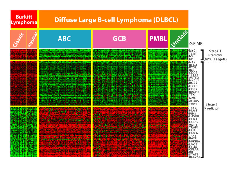 Lymphoma microarray.jpg 800 × 584; 178 KB
-
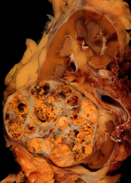 Renal cell ca.jpg 431 × 600; 70 KB
-
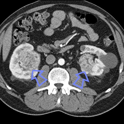 RCC.jpg 512 × 512; 55 KB
-
 Bonetumor.jpg 472 × 354; 73 KB
-
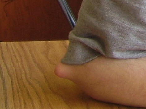
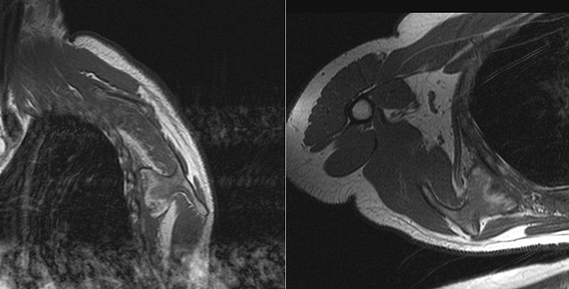 Osteochondroma right scapula.jpg 800 × 406; 44 KB
-
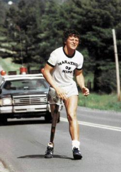 Terry fox running.jpg 250 × 356; 16 KB
-
 Canine osteosarcoma.JPG 800 × 600; 19 KB
-
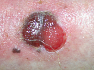
 Basaliom1.jpg 600 × 430; 146 KB
-
 Basaliom2.jpg 600 × 586; 185 KB
-
 Basaliom3.jpg 600 × 487; 166 KB
-
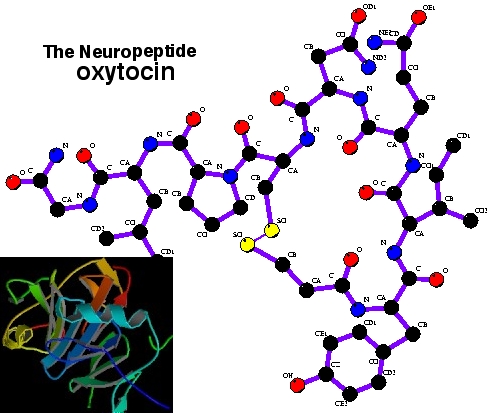 Oxytocin.jpg 493 × 413; 127 KB
-
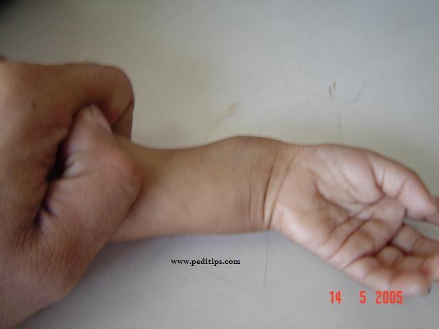 Rickewrist1.jpg 640 × 480; 23 KB
-
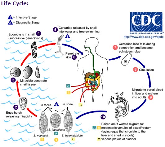 Life cycle schistosomiasis.jpg 532 × 476; 77 KB
-
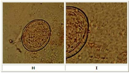 Schistosoma japonicum.jpg 421 × 239; 31 KB
-
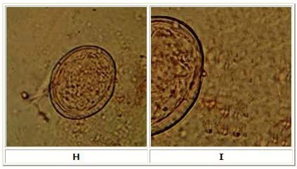 Schistosoma j.jpg 423 × 242; 32 KB
-
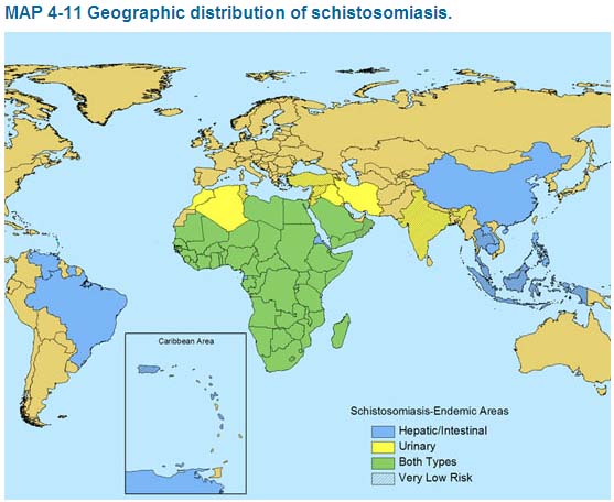 Map s.jpg 557 × 456; 63 KB
-
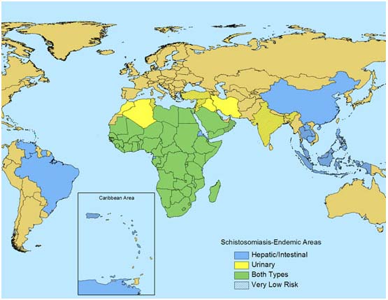 Map of distribution.jpg 555 × 429; 58 KB


.jpg)


.jpg)


_at_thigh_Case_01.jpg)
_at_thigh_Case_01.jpg)


