Liver abscess CT: Difference between revisions
Jump to navigation
Jump to search
No edit summary |
|||
| Line 10: | Line 10: | ||
Image:Liver-abscess-002.jpg|CT image demonstrates a large abscess in the right hepatic lobe | Image:Liver-abscess-002.jpg|CT image demonstrates a large abscess in the right hepatic lobe | ||
</gallery> | </gallery> | ||
Copyleft images obtained courtesy of RadsWiki [http://www.radswiki.net] | |||
Copyleft images obtained courtesy of RadsWiki [http://www.radswiki.net] | |||
==References== | ==References== | ||
Revision as of 17:50, 11 December 2012
|
Liver abscess Main Page |
Editor-In-Chief: C. Michael Gibson, M.S., M.D. [1]
CT Scan
On CT, abscesses may be single or multipe, round or oval, have an enhancing rim, complete or incomplete rim of edema, have smooth or nodular margins, intraabscess hemorrhage, peripheral biliary ductal dilatation, and may contain internal septations. Patterns are variable.
-
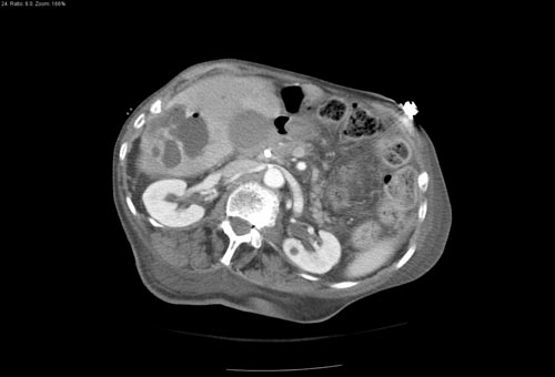
CT image demonstrates a large abscess in the right hepatic lobe
-
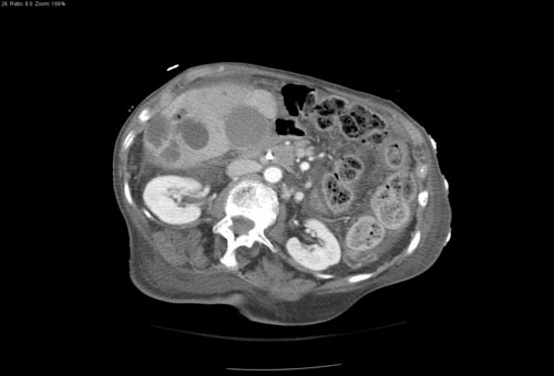
CT image demonstrates a large abscess in the right hepatic lobe


Copyleft images obtained courtesy of RadsWiki [2]