Torsades de pointes ekg examples: Difference between revisions
No edit summary |
No edit summary |
||
| Line 12: | Line 12: | ||
---- | ---- | ||
Shown below is an example of an ECG showing | Shown below is an example of an ECG showing [[wide QRS]] complexes in a twisted manner indicating torsades de pointes. | ||
[[image:Torsades de Pointes.1.4.1.jpg|center|800px]] | [[image:Torsades de Pointes.1.4.1.jpg|center|800px]] | ||
| Line 19: | Line 19: | ||
---- | ---- | ||
Shown below is an example of an ECG showing [[polymorphic ventricular tachycardia]] in a twisted appearance indicating torsades de pointes. | |||
[[image:Torsades de Pointes.1.4.2.jpg|center|800px]] | [[image:Torsades de Pointes.1.4.2.jpg|center|800px]] | ||
| Line 25: | Line 27: | ||
---- | ---- | ||
Shown below is an example of an ECG showing torsades de pointes. | |||
[[image:Torsades de Pointes.1.5.1.jpg|center|800px]] | [[image:Torsades de Pointes.1.5.1.jpg|center|800px]] | ||
Revision as of 16:24, 23 October 2012
Editor-In-Chief: C. Michael Gibson, M.S., M.D. [1]
EKG Examples
Shown below is an example of an ECG depicting wide QRS complexes in a twisted manner suggesting torsades de pointes.

Copyleft image obtained courtesy of ECGpedia, http://en.ecgpedia.org
Shown below is an example of an ECG showing wide QRS complexes in a twisted manner indicating torsades de pointes.

Copyleft image obtained courtesy of ECGpedia, http://en.ecgpedia.org
Shown below is an example of an ECG showing polymorphic ventricular tachycardia in a twisted appearance indicating torsades de pointes.

Copyleft image obtained courtesy of ECGpedia, http://en.ecgpedia.org
Shown below is an example of an ECG showing torsades de pointes.

Copyleft image obtained courtesy of ECGpedia, http://en.ecgpedia.org
Shown below is an example of an ECG showing arrhythmias in a patient with short coupled torsade de pointes[1]
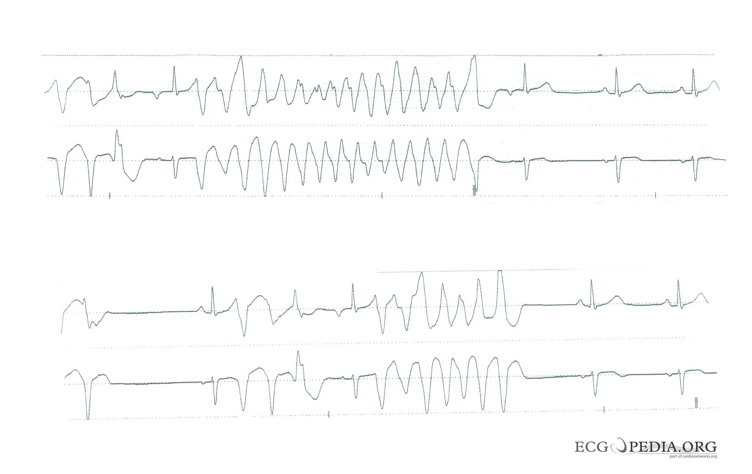
Copyleft image obtained courtesy of ECGpedia, http://en.ecgpedia.org
Shown below is an example of an ECG showing arrhythmias in a patient with short coupled torsades de pointes degenerating in ventricular fibrillation[1]
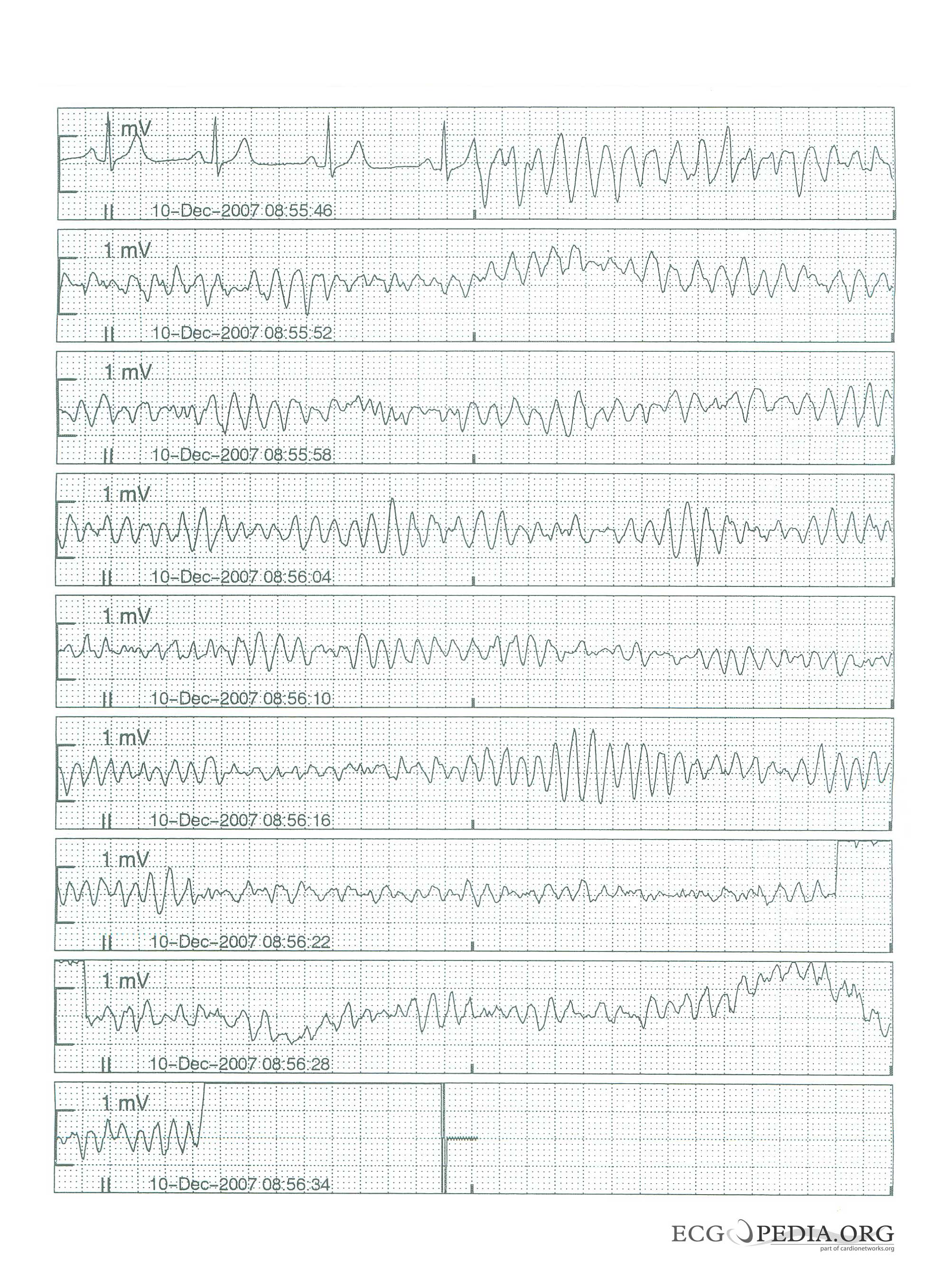
Copyleft image obtained courtesy of ECGpedia, http://en.ecgpedia.org
Shown below is an example of an ECG showing arrhythmias in a patient with short coupled torsade de pointes: frequent short coupled extrasystoles[1]
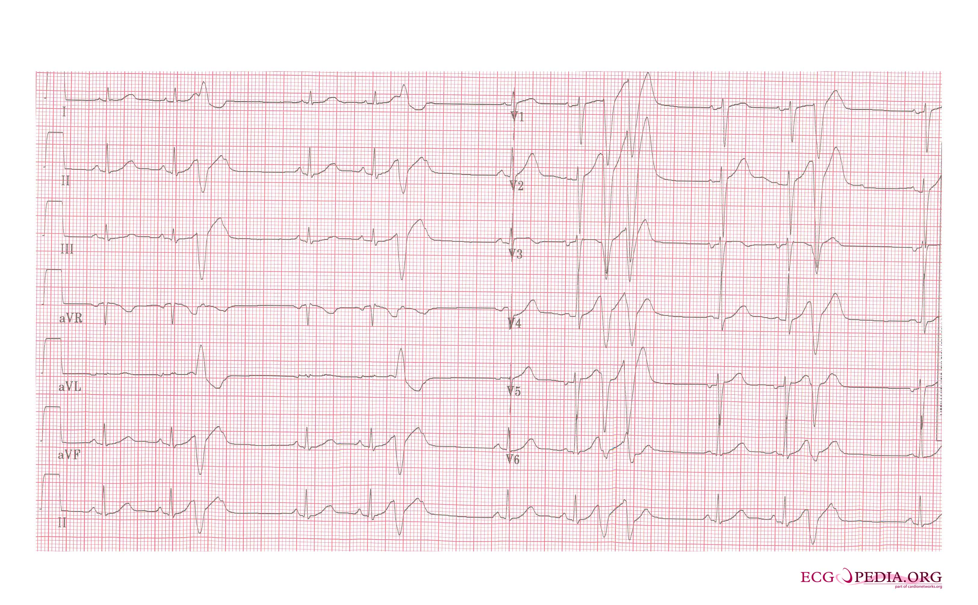
Copyleft image obtained courtesy of ECGpedia, http://en.ecgpedia.org
Shown below is an example of an ECG showing arrhythmias in a patient with short coupled torsade de pointes: frequent short coupled extrasystoles [1]
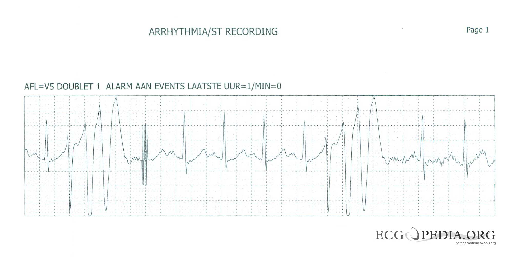
Copyleft image obtained courtesy of ECGpedia, http://en.ecgpedia.org
Shown below is an example of an ECG showing torsades de pointes[2]
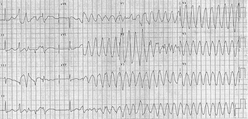
Copyleft image obtained courtesy of ECGpedia, http://en.ecgpedia.org
The ECG below is the characteristic tracing showing the "twisting" (blue line) of Torsade de pointes

Copyleft image obtained courtesy of Dr. C. Michael Gibson, MD.
References
- ↑ 1.0 1.1 1.2 1.3 Leenhardt A, Glaser E, Burguera M, Nuernberg M, Maison-Blanche P, and Coumel P. Short-coupled variant of torsade de pointes. A new electrocardiographic entity in the spectrum of idiopathic ventricular tachyarrhythmias. Circulation 1994 Jan; 89(1) 206-15. PMID 8281648
- ↑ Khan IA. Twelve-lead electrocardiogram of torsade de pointes Tex Heart Inst J. 2001; 28 (1): 69. PMID 11330748