Lipomatous tumor
Age of onset
Gender preponderance
Location
Clinical features
Pathologic appearance
Other features
Pathologic view
Angiolipoma
Second and third decades of life
More commonly seen in forearm
May also affect trunk and upper arm
Subcutaneous nodule
Tender to palpation
Less than 2 cm
Encapsulated, yellow nodules with a reddish tinge
A combination of fatty tissue and vascular channels
Fibrin thrombi is present in vascular channels (characteristic finding)
Contributed by Dr. Dharam Ramnani in Webpathology
Myolipoma
Fifth and sixth decades of life
More commonly seen in retroperitoneum, abdomen, pelvis, inguinal region, or abdominal wall
May also affect extremities
Subcutaneous mass which may also engage superficial muscular fascia
Size differs depending on the location
Partially encapsulated mass with partially yellow-white cut surface
A combination of mature adipocytes and sheets of well-differentiated smooth muscle
No nuclear atypia
Sieve-like appearance at low magnification (due to interspersed location of smooth muscle component)
Benign
It is usually large and located in the deep soft tissues
Contributed by Dr. Dharam Ramnani in Webpathology
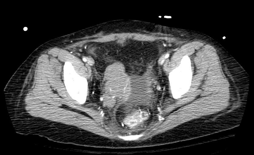
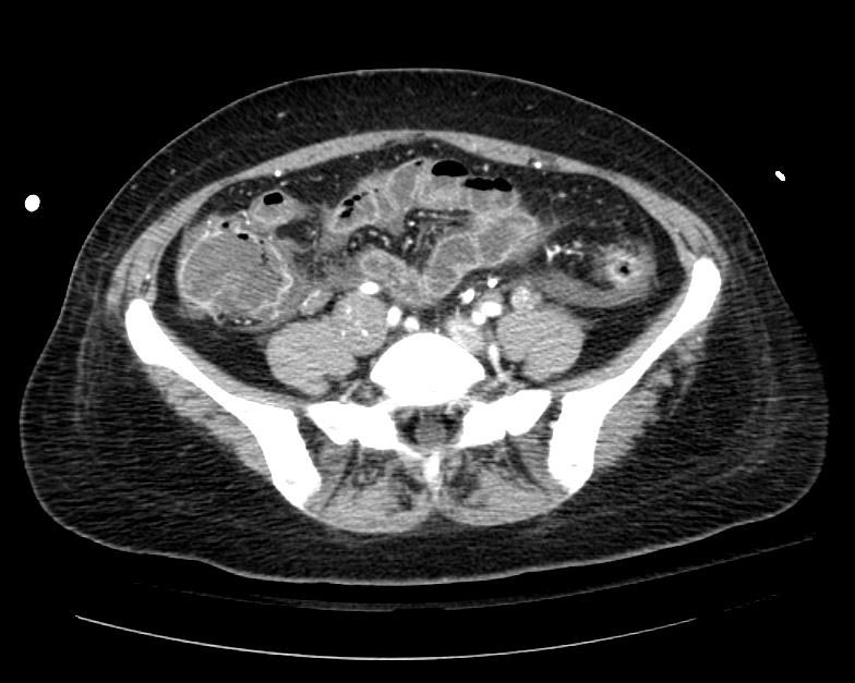
Myelolipoma
More commonly seen in adrenal glands
Other possible locations include:
thoracic, retroperitoneum and presacral region, mediastinum, liver, and bone
Usually asymptomatic
May cause abdominal pain, nausea, and constipation (depending on the location and size)
Uncommonly, may cause retro-peritoneal hemorrhage
3 to 7 cm
A combination of bone marrow elements and adipose tissue in varying proportions
May show myxoid changes
Well-circumscribed radiolucent mass in radiologic imaging
May have hromonal activity
Contributed by Sarahkayb in Wikimedia commons
Spindle Cell/Pleomorphic Lipoma
Fifth to seventh decades of life
More commonly seen in posterior neck, shoulder, and back
It is also reported in oral cavity
Subcutaneous nodule with firm consistency
Slowly growing and painless
Mostly between 3 to 5 cm
Similar to ordinary lipoma
A combination of mature fat cells and spindle cell or pleomorphic elements
Lipomatous component may vary in amount
Immunohistochemically positive for CD34
Benign
Contributed by Nephron in Wikimedia commons
Chondroid Lipoma
Third or fourth decade of life
More commonly seen in limbs and limb girdles
May also involve trunk, and the head and neck region, particularly the oral cavity
Slowly growing painless mass
Sizes ranges from 1 to 11 cm
Encapsulated tumor with a yellow, white, or pink-tan cut surface
A combination of mature adipocytes in association with nests of vacuolated cells in a myxochondroid or hyalinized fibrous background
Heterogeneous soft tissue mass in radiologic imaging
Benign
Contributed by Dr. Dharam Ramnani in Webpathology
Hibernoma
Most commonly seen in thigh
May also affect shoulder, back, neck, chest, arm, and abdominal cavity/retroperitoneum
Slowly growing, painless, subcutaneous mass
Affects intramuscular in 10% of the cases
Size varies between 5 to 15 cm
Well-defined, soft, and mobile mass
A combination of vacuolated granular eosinophilic cells with abundant mithochondria and high vascular content
Immunohistochemically positive for S-100
Benign
Contributed by Nephron in Wikimedia commons
Lipomas of Tendon Sheaths and Joints
Second and third decades of life
Intramuscular and Intermuscular Lipomas
Neural Fibrolipoma