Mesenteric ischemia CT: Difference between revisions
Feham Tariq (talk | contribs) (→CT) |
Feham Tariq (talk | contribs) No edit summary |
||
| Line 7: | Line 7: | ||
==CT== | ==CT== | ||
[[File:Venous-mesenteric-ischaemia-.jpg|400px|thumb|left|Red arrow shows Yellow arrows show ; Case courtesy of]] | |||
<br clear="left" /> | |||
Revision as of 18:44, 19 December 2017
|
Mesenteric ischemia Microchapters |
|
Diagnosis |
|---|
|
Treatment |
|
Case Studies |
|
Mesenteric ischemia CT On the Web |
|
American Roentgen Ray Society Images of Mesenteric ischemia CT |
|
Risk calculators and risk factors for Mesenteric ischemia CT |
Editor-In-Chief: C. Michael Gibson, M.S., M.D. [1] Associate Editor(s)-in-Chief: Feham Tariq, MD [2]
Overview
CT

Computed tomography (CT scan) is often used.[1][2] The accuracy of the CT scan depends on whether a small bowel obstruction (SBO) is present [3].
SBO absent
- prevalence of mesenteric ischemia 23%
- sensitivity 64%
- specificity 92%
- positive predictive value (at prevalence of 23%) 79%
- negative predictive value (at prevalence of 23%) 95%
SBO present
- prevalence of mesenteric ischemia 62%
- sensitivity 83%
- specificity 93%
- positive predictive value (at prevalence of 62%) 93%
- negative predictive value (at prevalence of 62%) 61%
Findings on CT scan include:
- Mesenteric edema[1]
- Bowel dilatation[1]
- Bowel wall thickening[1]
- Intramural gas[1]
- Mesenteric stranding[4]
- The CT and MR imaging findings represent a combination of those seen at plain radiography, barium studies, and angiography (vascular occlusion).
- CT and/or MR imaging may be helpful in determining the primary cause of bowel ischemia as well as allowing direct evaluation of the bowel wall, adjacent mesentery, and vascular structures.
- The most common CT finding in bowel ischemia is bowel wall thickening (nonspecific finding). The thickened bowel wall is sometimes associated with the target sign, alternating layers of high and low attenuation within the thickened bowel wall, which results from submucosal edema or hemorrhage.
- Absent or poor enhancement of the bowel wall is the most specific finding for bowel ischemia.
- Other CT findings of bowel ischemia reported in the literature include arterial occlusion, mesenteric or portal vein thrombosis, bowel dilatation, engorgement of mesenteric veins and mesenteric edema, intramural gas (intestinal pneumatosis), mesenteric or portal venous gas, lack of bowel wall enhancement, and infarction of other abdominal organs (eg, liver, spleen, or kidneys).
- Bowel dilatation reflects the interruption of peristaltic activity in ischemic segments. It is a common but nonspecific finding in bowel ischemia.
- Engorgement of mesenteric veins reflects venous congestion secondary to stasis.
- Owing to the edema that accompanies bowel ischemia, the mesenteric fat may be abnormally increased in attenuation.
- Intramural gas is a less common but more specific CT sign of ischemic bowel disease. The intramural gas is caused by dissection of luminal gas into the bowel wall across the compromised mucosa.
- Mesenteric or portal venous gas is an even less common CT manifestation of ischemic bowel disease and represents the propagation of intramural gas into the mesenteric venous system. Free intraperitoneal gas is an ominous CT sign in ischemic bowel disease because it indicates perforation of an infarcted bowel segment.
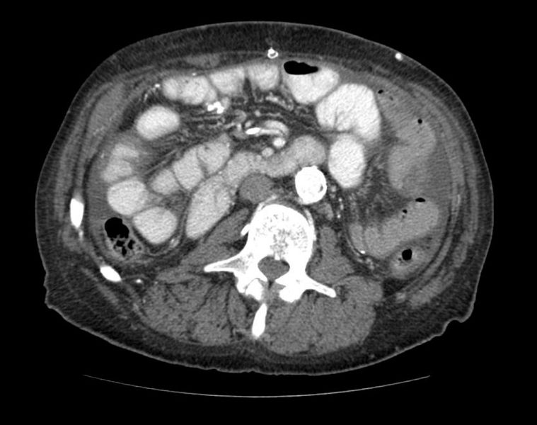
Patient #1: CT images of patient with ischemic bowel demonstrates pneumatosis and portal venous gas


Patient #2: CT images demonstrate mesenteric ischemia with marked atherosclerosis of arteries
-
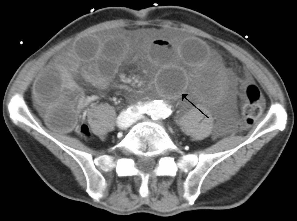
By James Heilman, MD - Own work, CC BY-SA 3.0, https://commons.wikimedia.org/w/index.php?curid=18076957
-
-

-

-

-

-


References
- ↑ 1.0 1.1 1.2 1.3 1.4 Alpern M, Glazer G, Francis I (1988). "Ischemic or infarcted bowel: CT findings". Radiology. 166 (1 Pt 1): 149–52. PMID 3336673.
- ↑ Taourel P, Deneuville M, Pradel J, Régent D, Bruel J (1996). "Acute mesenteric ischemia: diagnosis with contrast-enhanced CT" (PDF). Radiology. 199 (3): 632–6. doi:10.1148/rg.243035084. PMID 8637978.
- ↑ Staunton M, Malone DE (2005). "Can acute mesenteric ischemia be ruled out using computed tomography? Critically appraised topic". Canadian Association of Radiologists journal = Journal l'Association canadienne des radiologistes. 56 (1): 9–12. PMID 15835585.
- ↑ Pereira JM, Sirlin CB, Pinto PS, Jeffrey RB, Stella DL, Casola G (2004). "Disproportionate fat stranding: a helpful CT sign in patients with acute abdominal pain". Radiographics : a review publication of the Radiological Society of North America, Inc. 24 (3): 703–15. doi:10.1148/rg.243035084. PMID 15143223.