Sandbox 2: Difference between revisions
Jump to navigation
Jump to search
Aditya Ganti (talk | contribs) |
Aditya Ganti (talk | contribs) |
||
| Line 15: | Line 15: | ||
* The left colon stays perfused, primarily because of the marginal artery. | * The left colon stays perfused, primarily because of the marginal artery. | ||
{| border="1" cellpadding="5" cellspacing="0" align="center" |class="wikitable" | {| border="1" cellpadding="5" cellspacing="0" align="center" |class="wikitable" | ||
! colspan="2" align="center" style="background:#4479BA; color: #FFFFFF;" | ! colspan="2" align="center" style="background:#4479BA; color: #FFFFFF;" |Lower GI Tract | ||
!align="center" style="background:#4479BA; color: #FFFFFF;" | Arterial Supply | ! align="center" style="background:#4479BA; color: #FFFFFF;" | Arterial Supply | ||
!align="center" style="background:#4479BA; color: #FFFFFF;" |Venous Drainage | ! align="center" style="background:#4479BA; color: #FFFFFF;" |Venous Drainage | ||
|- | |- | ||
|style="padding: 5px 5px; background: #DCDCDC;" align="center" |Midgut | | style="padding: 5px 5px; background: #DCDCDC;" align="center" |Midgut | ||
|style="padding: 5px 5px; background: #F5F5F5;" align="left" | | | style="padding: 5px 5px; background: #F5F5F5;" align="left" | | ||
* Distal duodenum jejunum | * Distal duodenum jejunum | ||
* Ileum | * Ileum | ||
| Line 28: | Line 28: | ||
* Hepatic flexure | * Hepatic flexure | ||
* Proximal transverse colon. | * Proximal transverse colon. | ||
|style="padding: 5px 5px; background: #F5F5F5;" align="left" | | | style="padding: 5px 5px; background: #F5F5F5;" align="left" | | ||
* Superior mesenteric artery (SMA) | * Superior mesenteric artery (SMA) | ||
|style="padding: 5px 5px; background: #F5F5F5;" align="left" | | | style="padding: 5px 5px; background: #F5F5F5;" align="left" | | ||
* Portal system. | * Portal system. | ||
|- | |- | ||
|style="padding: 5px 5px; background: #DCDCDC;" align="center" |Hindgut | | style="padding: 5px 5px; background: #DCDCDC;" align="center" |Hindgut | ||
|style="padding: 5px 5px; background: #F5F5F5;" align="left" | | | style="padding: 5px 5px; background: #F5F5F5;" align="left" | | ||
* Distal one-third of the transverse colon | * Distal one-third of the transverse colon | ||
* Splenic flexure | * Splenic flexure | ||
| Line 40: | Line 40: | ||
* Sigmoid colon | * Sigmoid colon | ||
* Rectumhu | * Rectumhu | ||
|style="padding: 5px 5px; background: #F5F5F5;" align="left" | | | style="padding: 5px 5px; background: #F5F5F5;" align="left" | | ||
* Inferior mesenteric artery (IMA) | * Inferior mesenteric artery (IMA) | ||
|style="padding: 5px 5px; background: #F5F5F5;" align="left" | | | style="padding: 5px 5px; background: #F5F5F5;" align="left" | | ||
* Portal system '''<sup>ɸ</sup>''' | * Portal system '''<sup>ɸ</sup>''' | ||
|- | |- | ||
| colspan="4" style="padding: 5px 5px; background: #F5F5F5;" align="center" |ɸ -Except lower rectum, which drains into the systemic circulation. | | colspan="4" style="padding: 5px 5px; background: #F5F5F5;" align="center" |ɸ -Except lower rectum, which drains into the systemic circulation. | ||
|} | |} | ||
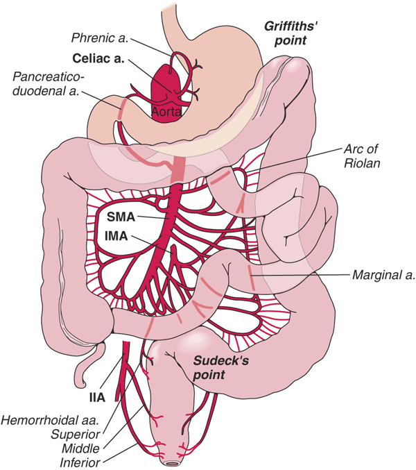
[[Image: Colonic blood supply1.gif|thumb|center|300px|Blood supply to the intestines includes the celiac artery, superior mesenteric artery (SMA), inferior mesenteric artery (IMA), and branches of the internal iliac artery (IIA). <br>Source: By Anpol42 (Own work) [CC BY-SA 4.0 (https://creativecommons.org/licenses/by-sa/4.0)], via Wikimedia Commons]] | [[Image: Colonic blood supply1.gif|thumb|center|300px|Blood supply to the intestines includes the celiac artery, superior mesenteric artery (SMA), inferior mesenteric artery (IMA), and branches of the internal iliac artery (IIA). <br>Source: By Anpol42 (Own work) [CC BY-SA 4.0 (https://creativecommons.org/licenses/by-sa/4.0)], via Wikimedia Commons]] | ||
| Line 54: | Line 52: | ||
===Pathogenesis=== | ===Pathogenesis=== | ||
Diverticulosis is the most common etiology of lower GI bleeding accounting for 30% of all cases, followed by anorectal disease, ischemia, inflammatory bowel disease (IBD), neoplasia and arteriovenous (AV) malformations. | Diverticulosis is the most common etiology of lower GI bleeding accounting for 30% of all cases, followed by anorectal disease, ischemia, inflammatory bowel disease (IBD), neoplasia and arteriovenous (AV) malformations. | ||
*'''Diverticulosis''' | *'''<u>Diverticulosis</u>''' | ||
:*The colonic wall weakens with age and results in the formation of saclike protrusions known as diverticula. | :*The colonic wall weakens with age and results in the formation of saclike protrusions known as diverticula. | ||
:*These protrusions generally occur at the junction of blood vessel penetrating through the mucosa and circular muscle fibers of the colon. | :*These protrusions generally occur at the junction of blood vessel penetrating through the mucosa and circular muscle fibers of the colon. | ||
| Line 61: | Line 59: | ||
:*Most of the time bleeding from diverticulosis stops spontaneously, however, in about 5% of patients, the bleeding can be massive and life-threatening. | :*Most of the time bleeding from diverticulosis stops spontaneously, however, in about 5% of patients, the bleeding can be massive and life-threatening. | ||
[[Image:Sigmoid diverticulum (diagram).jpg|thumb|center|400px|Diagram of sigmoid diverticulum<br>Source:By Anpol42 (Own work) [CC BY-SA 4.0 (https://creativecommons.org/licenses/by-sa/4.0)], via Wikimedia Commons]] | [[Image:Sigmoid diverticulum (diagram).jpg|thumb|center|400px|Diagram of sigmoid diverticulum<br>Source:By Anpol42 (Own work) [CC BY-SA 4.0 (https://creativecommons.org/licenses/by-sa/4.0)], via Wikimedia Commons]] | ||
*'''Anorectal disease''' | *'''<u>Anorectal disease</u>''' | ||
*: Hemorrhoids and anal fissures are the most common disease under anorectal disease responsible for GI bleeding. | *: Hemorrhoids and anal fissures are the most common disease under anorectal disease responsible for GI bleeding. | ||
:*Hemorrhoids are engorged vessels in the normal anal cushions. When swollen, this tissue is very friable and susceptible to trauma, which leads to painless, bright red bleeding. | :*Hemorrhoids are engorged vessels in the normal anal cushions. When swollen, this tissue is very friable and susceptible to trauma, which leads to painless, bright red bleeding. | ||
:*Anal fissures are defined as a tear in the anal mucosa. With the passage of stool, the mucosa continues to tear and leads to bright red bleeding. | :*Anal fissures are defined as a tear in the anal mucosa. With the passage of stool, the mucosa continues to tear and leads to bright red bleeding. | ||
*'''Mesenteric Ischemia''' | *'''<u>Mesenteric Ischemia</u>''' | ||
Revision as of 19:33, 20 November 2017
Lower GI bleeding is defined as any bleed that occurs distal to the ligament of Treitz.
Incidence
- In the United States the incidence of LGIB ranges from 20.5 to 27 per 100,000 persons per year.
Age
- There is a greater than 200 fold increase from the third to the ninth decade of life.
Classification
- Lower GI bleeding can be classified into 3 groups based on the severity of bleeding:
- Occult lower GI bleeding
- Moderate lower GI bleeding
- Severe lower GI bleeding
Blood supply
- The SMA and IMA are connected by the marginal artery of Drummond.
- This vascular arcade runs in the mesentery close to the bowel.
- As patients age, there is increased incidence of occlusion of the IMA.
- The left colon stays perfused, primarily because of the marginal artery.
| Lower GI Tract | Arterial Supply | Venous Drainage | |
|---|---|---|---|
| Midgut |
|
|
|
| Hindgut |
|
|
|
| ɸ -Except lower rectum, which drains into the systemic circulation. | |||

Source: By Anpol42 (Own work) [CC BY-SA 4.0 (https://creativecommons.org/licenses/by-sa/4.0)], via Wikimedia Commons
Pathogenesis
Diverticulosis is the most common etiology of lower GI bleeding accounting for 30% of all cases, followed by anorectal disease, ischemia, inflammatory bowel disease (IBD), neoplasia and arteriovenous (AV) malformations.
- Diverticulosis
- The colonic wall weakens with age and results in the formation of saclike protrusions known as diverticula.
- These protrusions generally occur at the junction of blood vessel penetrating through the mucosa and circular muscle fibers of the colon.
- Diverticula are most common in the descending and sigmoid colon.
- Despite the majority of diverticula being on the left side of the colon, diverticular bleeding originates from the right side of the colon in 50% to 90% of instances.
- Most of the time bleeding from diverticulosis stops spontaneously, however, in about 5% of patients, the bleeding can be massive and life-threatening.

Source:By Anpol42 (Own work) [CC BY-SA 4.0 (https://creativecommons.org/licenses/by-sa/4.0)], via Wikimedia Commons
- Anorectal disease
- Hemorrhoids and anal fissures are the most common disease under anorectal disease responsible for GI bleeding.
- Hemorrhoids are engorged vessels in the normal anal cushions. When swollen, this tissue is very friable and susceptible to trauma, which leads to painless, bright red bleeding.
- Anal fissures are defined as a tear in the anal mucosa. With the passage of stool, the mucosa continues to tear and leads to bright red bleeding.
- Mesenteric Ischemia