Hepatic hemangioma pathophysiology: Difference between revisions
Nawal Muazam (talk | contribs) No edit summary |
Nawal Muazam (talk | contribs) No edit summary |
||
| Line 4: | Line 4: | ||
==Overview== | ==Overview== | ||
==Pathophysiology== | ==Pathophysiology== | ||
===Pathogenesis=== | |||
* They arise from the endothelial cells that line the blood vessels and consists of multiple, large vascular channels lined by a single layer of endothelial cells and supported by collagenous walls. | * They arise from the endothelial cells that line the blood vessels and consists of multiple, large vascular channels lined by a single layer of endothelial cells and supported by collagenous walls. | ||
* They may be associated with [[focal nodular hyperplasia]]. | * They may be associated with [[focal nodular hyperplasia]]. | ||
===Associated Conditions=== | ===Associated Conditions=== | ||
* May be associated with [[Kasabach-Merritt syndrome]], [[hemolytic anemia]] and consumptive coagulopathy. | * May be associated with [[Kasabach-Merritt syndrome]], [[hemolytic anemia]] and consumptive coagulopathy. | ||
==Gross Pathology== | ===Gross Pathology=== | ||
On gross pathology, variable in size, well circumscribed, classically subcapsular are characteristic findings of hepatic hemangioma.<ref name=bc>Gross pathology of hepatic hemangioma. Librepathology 2015. http://librepathology.org/wiki/index.php/Hemangioma_of_the_liver. Accessed on October 20, 2015</ref> | On gross pathology, variable in size, well circumscribed, classically subcapsular are characteristic findings of hepatic hemangioma.<ref name=bc>Gross pathology of hepatic hemangioma. Librepathology 2015. http://librepathology.org/wiki/index.php/Hemangioma_of_the_liver. Accessed on October 20, 2015</ref> | ||
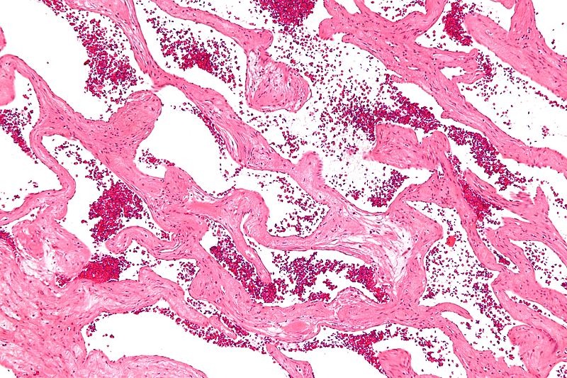
==Microscopic Pathology== | ===Microscopic Pathology=== | ||
On microscopic histopathological analysis channels lined by benign endothelium containing RBCs, surrounding (non-endothelial) cells without significant atypia are characteristic findings of hepatic hemangioma.<ref name=bc>Microscopic features of hepatic hemangioma. Librepathology 2015. http://librepathology.org/wiki/index.php/Hemangioma_of_the_liver. Accessed on October 20, 2015</ref> | On microscopic histopathological analysis channels lined by benign endothelium containing RBCs, surrounding (non-endothelial) cells without significant atypia are characteristic findings of hepatic hemangioma.<ref name=bc>Microscopic features of hepatic hemangioma. Librepathology 2015. http://librepathology.org/wiki/index.php/Hemangioma_of_the_liver. Accessed on October 20, 2015</ref> | ||
====Gallery==== | |||
<gallery> | |||
Image:Cavernous_liver_hemangioma_-_intermed_mag.jpg|Intermediate magnification micrograph of a cavernous hemangioma of the liver, also hepatic cavernous hemangioma, liver hemangioma,cavernous liver hemangioma. H&E stain. No liver tissue is observed. | |||
</gallery> | |||
Revision as of 20:07, 20 October 2015
|
Hepatic hemangioma Microchapters |
|
Diagnosis |
|---|
|
Treatment |
|
Case Studies |
|
Hepatic hemangioma pathophysiology On the Web |
|
American Roentgen Ray Society Images of Hepatic hemangioma pathophysiology |
|
Risk calculators and risk factors for Hepatic hemangioma pathophysiology |
Editor-In-Chief: C. Michael Gibson, M.S., M.D. [1];Associate Editor(s)-in-Chief: Nawal Muazam M.D.[2]
Overview
Pathophysiology
Pathogenesis
- They arise from the endothelial cells that line the blood vessels and consists of multiple, large vascular channels lined by a single layer of endothelial cells and supported by collagenous walls.
- They may be associated with focal nodular hyperplasia.
Associated Conditions
- May be associated with Kasabach-Merritt syndrome, hemolytic anemia and consumptive coagulopathy.
Gross Pathology
On gross pathology, variable in size, well circumscribed, classically subcapsular are characteristic findings of hepatic hemangioma.[1]
Microscopic Pathology
On microscopic histopathological analysis channels lined by benign endothelium containing RBCs, surrounding (non-endothelial) cells without significant atypia are characteristic findings of hepatic hemangioma.[1]
Gallery
-
Intermediate magnification micrograph of a cavernous hemangioma of the liver, also hepatic cavernous hemangioma, liver hemangioma,cavernous liver hemangioma. H&E stain. No liver tissue is observed.

References
- ↑ 1.0 1.1 Gross pathology of hepatic hemangioma. Librepathology 2015. http://librepathology.org/wiki/index.php/Hemangioma_of_the_liver. Accessed on October 20, 2015