Paget-Schroetter disease echocardiography or ultrasound: Difference between revisions
No edit summary |
No edit summary |
||
| (6 intermediate revisions by 2 users not shown) | |||
| Line 1: | Line 1: | ||
__NOTOC__ | __NOTOC__ | ||
{{Paget-Schroetter disease}} | |||
{{CMG}}; {{AE}} {{Anahita}} | |||
== Overview == | == Overview == | ||
[[Paget-Schroetter disease]] is commonly diagnosed with a history and [[Physical examination|physical examinations]]. However, imaging is usually utilized to confirm the diagnose. [[Duplex ultrasound]] is an accepted initial test and the [[Gold standard (test)|gold standard]] imaging of [[Paget-Schroetter disease]]. Since this diagnostic tool is not fully appropriate to exclude the Paget-Schroetter disease, normal [[duplex ultrasound]] in a highly suspected patient requires further investigations. | |||
== Ultrasound == | == Ultrasound == | ||
| Line 15: | Line 17: | ||
** [[Sensitivity (tests)|Sensitivity]] of >80%<ref name="Rosa SalazarOtálora Valderrama20152">{{cite journal|last1=Rosa Salazar|first1=Vladimir|last2=Otálora Valderrama|first2=Sonia del Pilar|last3=Hernández Contreras|first3=María Encarnación|last4=García Pérez|first4=Bartolomé|last5=Arroyo Tristán|first5=Andrés del Amor|last6=García Méndez|first6=María del Mar|title=Multidisciplinary Management of Paget-Schroetter Syndrome. A Case Series of Eight Patients|journal=Archivos de Bronconeumología (English Edition)|volume=51|issue=8|year=2015|pages=e41–e43|issn=15792129|doi=10.1016/j.arbr.2015.05.026}}</ref> | ** [[Sensitivity (tests)|Sensitivity]] of >80%<ref name="Rosa SalazarOtálora Valderrama20152">{{cite journal|last1=Rosa Salazar|first1=Vladimir|last2=Otálora Valderrama|first2=Sonia del Pilar|last3=Hernández Contreras|first3=María Encarnación|last4=García Pérez|first4=Bartolomé|last5=Arroyo Tristán|first5=Andrés del Amor|last6=García Méndez|first6=María del Mar|title=Multidisciplinary Management of Paget-Schroetter Syndrome. A Case Series of Eight Patients|journal=Archivos de Bronconeumología (English Edition)|volume=51|issue=8|year=2015|pages=e41–e43|issn=15792129|doi=10.1016/j.arbr.2015.05.026}}</ref> | ||
** Non-invasive | ** Non-invasive | ||
* Based on some [[Randomized controlled trial|randomized trial]] [[duplex ultrasound]] has a [[Sensitivity (tests)|sensitivity]] of 78-100% and [[Specificity (tests)| | * Based on some [[Randomized controlled trial|randomized trial]] [[duplex ultrasound]] has a [[Sensitivity (tests)|sensitivity]] of 78-100% and [[Specificity (tests)|specificity]] of 82-100%. <ref name="IlligDoyle2010">{{cite journal|last1=Illig|first1=Karl A.|last2=Doyle|first2=Adam J.|title=A comprehensive review of Paget-Schroetter syndrome|journal=Journal of Vascular Surgery|volume=51|issue=6|year=2010|pages=1538–1547|issn=07415214|doi=10.1016/j.jvs.2009.12.022}}</ref> | ||
* An experienced technician is able to distinguish between [[Acute (medicine)|acute]] and [[Chronic (medical)|chronic]] [[thrombus]] using [[ | * An experienced technician is able to distinguish between [[Acute (medicine)|acute]] and [[Chronic (medical)|chronic]] [[thrombus]] using [[duplex ultrasound]]. | ||
*An [[Acute (medicine)|acute]] [[thrombus]] which is echolucent leads to a non-compressible vein without any flow. In the contrary, a [[Chronic (medical)|chronic]] [[thrombus]] will appear [[Echogenicity|echogenic]] and fibrotic with notable collateral veins. <ref name="IlligDoyle20103">{{cite journal|last1=Illig|first1=Karl A.|last2=Doyle|first2=Adam J.|title=A comprehensive review of Paget-Schroetter syndrome|journal=Journal of Vascular Surgery|volume=51|issue=6|year=2010|pages=1538–1547|issn=07415214|doi=10.1016/j.jvs.2009.12.022}}</ref> | |||
* Some drawbacks of [[Duplex ultrasound|Duplex ultrasonography]] are: | * Some drawbacks of [[Duplex ultrasound|Duplex ultrasonography]] are: | ||
** Inadequate ability to picture the central parts of the [[ | ** Inadequate ability to picture the central parts of the [[subclavian]] and [[innominate]] veins <ref name="IlligDoyle20104">{{cite journal|last1=Illig|first1=Karl A.|last2=Doyle|first2=Adam J.|title=A comprehensive review of Paget-Schroetter syndrome|journal=Journal of Vascular Surgery|volume=51|issue=6|year=2010|pages=1538–1547|issn=07415214|doi=10.1016/j.jvs.2009.12.022}}</ref> | ||
** Inadequate ability to distinguish central veins from an enlarged collateral vein<ref name="IlligDoyle20105">{{cite journal|last1=Illig|first1=Karl A.|last2=Doyle|first2=Adam J.|title=A comprehensive review of Paget-Schroetter syndrome|journal=Journal of Vascular Surgery|volume=51|issue=6|year=2010|pages=1538–1547|issn=07415214|doi=10.1016/j.jvs.2009.12.022}}</ref> | ** Inadequate ability to distinguish central veins from an enlarged collateral vein<ref name="IlligDoyle20105">{{cite journal|last1=Illig|first1=Karl A.|last2=Doyle|first2=Adam J.|title=A comprehensive review of Paget-Schroetter syndrome|journal=Journal of Vascular Surgery|volume=51|issue=6|year=2010|pages=1538–1547|issn=07415214|doi=10.1016/j.jvs.2009.12.022}}</ref> | ||
** Highly technician dependent<ref name="Thompson20122">{{cite journal|last1=Thompson|first1=Robert|title=Comprehensive Management of Subclavian Vein Effort Thrombosis|journal=Seminars in Interventional Radiology|volume=29|issue=01|year=2012|pages=044–051|issn=0739-9529|doi=10.1055/s-0032-1302451}}</ref> | ** Highly technician dependent<ref name="Thompson20122">{{cite journal|last1=Thompson|first1=Robert|title=Comprehensive Management of Subclavian Vein Effort Thrombosis|journal=Seminars in Interventional Radiology|volume=29|issue=01|year=2012|pages=044–051|issn=0739-9529|doi=10.1055/s-0032-1302451}}</ref> | ||
** Findings could be affected by superimposed [[clavicle]] and deepness of | ** Findings could be affected by superimposed [[clavicle]] and deepness of subclavian vein in the neck<ref name="Thompson2012">{{cite journal|last1=Thompson|first1=Robert|title=Comprehensive Management of Subclavian Vein Effort Thrombosis|journal=Seminars in Interventional Radiology|volume=29|issue=01|year=2012|pages=044–051|issn=0739-9529|doi=10.1055/s-0032-1302451}}</ref> | ||
** [[False negative|False negative rate]] of 30% (which makes [[Duplex ultrasound| | **[[False negative|False negative rate]] of 30% (which makes [[Duplex ultrasound|duplex ultrasonography]] an improper diagnostic tool to exclude the [[Paget-Schroetter disease]])<ref name="Thompson20123">{{cite journal|last1=Thompson|first1=Robert|title=Comprehensive Management of Subclavian Vein Effort Thrombosis|journal=Seminars in Interventional Radiology|volume=29|issue=01|year=2012|pages=044–051|issn=0739-9529|doi=10.1055/s-0032-1302451}}</ref> | ||
* [[Ultrasound]] as a non-invasive imaging could be used postoperatively to evaluate venous flow and caliber.<ref name="Rubin2009">{{cite journal|last1=Rubin|first1=Joseph|title=Invited Commentary|journal=The Annals of Thoracic Surgery|volume=87|issue=2|year=2009|pages=422|issn=00034975|doi=10.1016/j.athoracsur.2008.11.058}}</ref> | *[[Ultrasound]] as a non-invasive imaging could be used postoperatively to evaluate venous flow and caliber.<ref name="Rubin2009">{{cite journal|last1=Rubin|first1=Joseph|title=Invited Commentary|journal=The Annals of Thoracic Surgery|volume=87|issue=2|year=2009|pages=422|issn=00034975|doi=10.1016/j.athoracsur.2008.11.058}}</ref> | ||
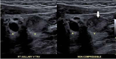
[[File:Noncompressive Axillary vein.jpg|alt=Noncompressive Axillary vein |center|thumb|398x398px|Doppler ultrasound showing noncompressibility of the axillary vein (arrow) due to thrombus. Case courtesy by Himani Sharma, MD<ref>{{Cite web|url=https://www.hindawi.com/journals/crivam/2017/8764903/|title=Recurrent Upper Extremity Thrombosis Associated with Overactivity: A Case of Delayed Diagnosis of Paget-Schroetter Syndrome|last=|first=|date=|website=|archive-url=|archive-date=|dead-url=|access-date=}}</ref>]] | |||
<br /> | |||
==References== | ==References== | ||
{{Reflist|2}} | {{Reflist|2}} | ||
[[Category:Hematology]] | [[Category:Hematology]] | ||
[[Category:Angiology]] | [[Category:Angiology]] | ||
[[Category:cardiology]] | [[Category:cardiology]] | ||
{{WH}} | {{WH}} | ||
{{WS}} | {{WS}} | ||
Latest revision as of 20:19, 23 June 2020
|
Paget-Schroetter disease Microchapters |
|
Differentiating Paget-Schroetter disease from other Diseases |
|---|
|
Diagnosis |
|
Treatment |
|
Case Studies |
|
Paget-Schroetter disease echocardiography or ultrasound On the Web |
|
American Roentgen Ray Society Images of Paget-Schroetter disease echocardiography or ultrasound |
|
FDA on Paget-Schroetter disease echocardiography or ultrasound |
|
Paget-Schroetter disease echocardiography or ultrasound in the news |
|
Blogs on Paget-Schroetter disease echocardiography or ultrasound |
|
Risk calculators and risk factors for Paget-Schroetter disease echocardiography or ultrasound |
Editor-In-Chief: C. Michael Gibson, M.S., M.D. [1]; Associate Editor(s)-in-Chief: Anahita Deylamsalehi, M.D.[2]
Overview
Paget-Schroetter disease is commonly diagnosed with a history and physical examinations. However, imaging is usually utilized to confirm the diagnose. Duplex ultrasound is an accepted initial test and the gold standard imaging of Paget-Schroetter disease. Since this diagnostic tool is not fully appropriate to exclude the Paget-Schroetter disease, normal duplex ultrasound in a highly suspected patient requires further investigations.
Ultrasound
- Duplex ultrasound is the gold standard method to diagnose Paget-Schroetter (effort thrombosis).[1]
- Doppler ultrasonography is the best initial test to evaluate a patient with suspected Paget-Schroetter disease (also known as Effort thrombosis).[2] There are some convenient features of this test that make it a proper initial imaging. These features include:
- Low cost [3]
- Availability
- Portability
- Specificity of 93%[4]
- Sensitivity of >80%[5]
- Non-invasive
- Based on some randomized trial duplex ultrasound has a sensitivity of 78-100% and specificity of 82-100%. [6]
- An experienced technician is able to distinguish between acute and chronic thrombus using duplex ultrasound.
- An acute thrombus which is echolucent leads to a non-compressible vein without any flow. In the contrary, a chronic thrombus will appear echogenic and fibrotic with notable collateral veins. [7]
- Some drawbacks of Duplex ultrasonography are:
- Inadequate ability to picture the central parts of the subclavian and innominate veins [8]
- Inadequate ability to distinguish central veins from an enlarged collateral vein[9]
- Highly technician dependent[10]
- Findings could be affected by superimposed clavicle and deepness of subclavian vein in the neck[11]
- False negative rate of 30% (which makes duplex ultrasonography an improper diagnostic tool to exclude the Paget-Schroetter disease)[12]
- Ultrasound as a non-invasive imaging could be used postoperatively to evaluate venous flow and caliber.[13]

References
- ↑ Hangge, Patrick; Rotellini-Coltvet, Lisa; Deipolyi, Amy R.; Albadawi, Hassan; Oklu, Rahmi (2017). "Paget-Schroetter syndrome: treatment of venous thrombosis and outcomes". Cardiovascular Diagnosis and Therapy. 7 (S3): S285–S290. doi:10.21037/cdt.2017.08.15. ISSN 2223-3652.
- ↑ Alla VM, Natarajan N, Kaushik M, Warrier R, Nair CK (2010). "Paget-schroetter syndrome: review of pathogenesis and treatment of effort thrombosis". West J Emerg Med. 11 (4): 358–62. PMC 2967689. PMID 21079709.
- ↑ Alla VM, Natarajan N, Kaushik M, Warrier R, Nair CK (2010). "Paget-schroetter syndrome: review of pathogenesis and treatment of effort thrombosis". West J Emerg Med. 11 (4): 358–62. PMC 2967689. PMID 21079709.
- ↑ Rosa Salazar, Vladimir; Otálora Valderrama, Sonia del Pilar; Hernández Contreras, María Encarnación; García Pérez, Bartolomé; Arroyo Tristán, Andrés del Amor; García Méndez, María del Mar (2015). "Multidisciplinary Management of Paget-Schroetter Syndrome. A Case Series of Eight Patients". Archivos de Bronconeumología (English Edition). 51 (8): e41–e43. doi:10.1016/j.arbr.2015.05.026. ISSN 1579-2129.
- ↑ Rosa Salazar, Vladimir; Otálora Valderrama, Sonia del Pilar; Hernández Contreras, María Encarnación; García Pérez, Bartolomé; Arroyo Tristán, Andrés del Amor; García Méndez, María del Mar (2015). "Multidisciplinary Management of Paget-Schroetter Syndrome. A Case Series of Eight Patients". Archivos de Bronconeumología (English Edition). 51 (8): e41–e43. doi:10.1016/j.arbr.2015.05.026. ISSN 1579-2129.
- ↑ Illig, Karl A.; Doyle, Adam J. (2010). "A comprehensive review of Paget-Schroetter syndrome". Journal of Vascular Surgery. 51 (6): 1538–1547. doi:10.1016/j.jvs.2009.12.022. ISSN 0741-5214.
- ↑ Illig, Karl A.; Doyle, Adam J. (2010). "A comprehensive review of Paget-Schroetter syndrome". Journal of Vascular Surgery. 51 (6): 1538–1547. doi:10.1016/j.jvs.2009.12.022. ISSN 0741-5214.
- ↑ Illig, Karl A.; Doyle, Adam J. (2010). "A comprehensive review of Paget-Schroetter syndrome". Journal of Vascular Surgery. 51 (6): 1538–1547. doi:10.1016/j.jvs.2009.12.022. ISSN 0741-5214.
- ↑ Illig, Karl A.; Doyle, Adam J. (2010). "A comprehensive review of Paget-Schroetter syndrome". Journal of Vascular Surgery. 51 (6): 1538–1547. doi:10.1016/j.jvs.2009.12.022. ISSN 0741-5214.
- ↑ Thompson, Robert (2012). "Comprehensive Management of Subclavian Vein Effort Thrombosis". Seminars in Interventional Radiology. 29 (01): 044–051. doi:10.1055/s-0032-1302451. ISSN 0739-9529.
- ↑ Thompson, Robert (2012). "Comprehensive Management of Subclavian Vein Effort Thrombosis". Seminars in Interventional Radiology. 29 (01): 044–051. doi:10.1055/s-0032-1302451. ISSN 0739-9529.
- ↑ Thompson, Robert (2012). "Comprehensive Management of Subclavian Vein Effort Thrombosis". Seminars in Interventional Radiology. 29 (01): 044–051. doi:10.1055/s-0032-1302451. ISSN 0739-9529.
- ↑ Rubin, Joseph (2009). "Invited Commentary". The Annals of Thoracic Surgery. 87 (2): 422. doi:10.1016/j.athoracsur.2008.11.058. ISSN 0003-4975.
- ↑ "Recurrent Upper Extremity Thrombosis Associated with Overactivity: A Case of Delayed Diagnosis of Paget-Schroetter Syndrome".