Trigeminal nerve nuclei
(Redirected from Trigeminal nucleus)
Overview
The sensory trigeminal nerve nucleus is the largest of the cranial nerve nuclei, and extends through the whole of the brainstem, midbrain to medulla.
The nucleus is divided into three parts, from rostral to caudal (top to bottom in humans):
- The mesencephalic nucleus
- The chief sensory nucleus (or "pontine nucleus")
- The spinal trigeminal nucleus
There is also a distinct trigeminal motor nucleus that is medial to the chief sensory nucleus.
See also
Additional images
-

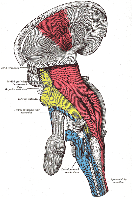
Dissection of brain-stem. Lateral view.
-
Deep dissection of brain-stem. Lateral view.
-
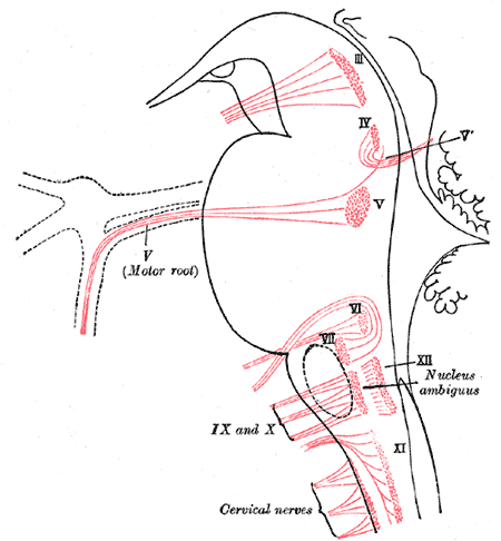
Nuclei of origin of cranial motor nerves schematically represented; lateral view.
-

Primary terminal nuclei of the afferent (sensory) cranial nerves schematically represented; lateral view.


