The heart in polyarteritis nodosa
You don't need to be Editor-In-Chief to add or edit content to WikiDoc. You can begin to add to or edit text on this WikiDoc page by clicking on the edit button at the top of this page. Next enter or edit the information that you would like to appear here. Once you are done editing, scroll down and click the Save page button at the bottom of the page.
| The heart in polyarteritis nodosa Classification and external resources | |
| Image courtesy of Professor Peter Anderson DVM PhD and published with permission © PEIR, University of Alabama at Birmingham, Department of Pathology |
Editor-In-Chief: C. Michael Gibson, M.S., M.D. [1] Phone:617-525-6884
Associate Editor: Cafer Zorkun, M.D., Ph.D. [2] Phone:617-525-7431
| Cardiology Network |
| Discuss The heart in polyarteritis nodosa further in the WikiDoc Cardiology Network |
| Adult Congenital |
|---|
| Biomarkers |
| Cardiac Rehabilitation |
| Congestive Heart Failure |
| CT Angiography |
| Echocardiography |
| Electrophysiology |
| Cardiology General |
| Genetics |
| Health Economics |
| Hypertension |
| Interventional Cardiology |
| MRI |
| Nuclear Cardiology |
| Peripheral Arterial Disease |
| Prevention |
| Public Policy |
| Pulmonary Embolism |
| Stable Angina |
| Valvular Heart Disease |
| Vascular Medicine |
| WikiDoc Cardiology News |
![]() Read more about The heart in polyarteritis nodosa in the WikiDoc Cardiology News |
| All News Articles |
|---|
| Acute Coronary Syndromes |
| Biomarkers |
| Cardiovascular Imaging |
| CT Surgery |
| Diabetes |
| Electrophysiology |
| General Cardiology |
| Guidelines |
| Health Policy |
| Heart Failure |
| Hypertension |
| Interventional |
| Peripheral Arterial Disease |
| Prevention |
| Valvular Heart Disease |
Please Take Over This Page and Apply to be Editor-In-Chief for this topic: There can be one or more than one Editor-In-Chief. You may also apply to be an Associate Editor-In-Chief of one of the subtopics below. Please mail us [3] to indicate your interest in serving either as an Editor-In-Chief of the entire topic or as an Associate Editor-In-Chief for a subtopic. Please be sure to attach your CV and or biographical sketch.
The heart and coronary arteries are infrequently involved in polyarteritis nodosa. Most often this involvement is a vasculitis of the distal subepicardial coronary arteries just as they penetrate the myocardium. The lesions are characterized by inflammatory infiltrates in the media and adventitia and occasionally by necrosis of the full thickness of the vessel wall, with prominent involvement of the surrounding perivascular connective tissue.
The lumens of the involved vessels may contain thrombi, and the walls may be aneurysmal. The latter is responsible for the nodular appearance of the arteries deemed characteristic of this disorder. An even later stage of the vasculitis process is evident as the lesions heal, first showing the formation of granulation tissue and subsequently fibrous tissue replacement of the original components of the artery. In this healing phase, intimal proliferation leading to coronary artery luminal narrowing is evident.
The coronary artery disease of Polyarteritis Nodosa may lead to myocardial infarction. The myocardial necrosis and subsequent replacement fibrosis tend to be focal and patchy throughout the left ventricular wall. This is in contrast to the large areas of grossly visible, regional, subendocardial, or transmural necrosis typically seen in the myocardial infarction caused by Coronary Artery Disease.
Conduction system abnormalities have been identified in the heart of patients with Polyarteritis Nodosa. The size and location of the sinoatrial node and atrioventricular node arteries make them prime targets for polyarteritis. Atrial and ventricular conduction disturbances may be a primary manifestation of Polyarteritis Nodosa, despite minimal involvement of vessels elsewhere in the heart.
Other cardiac abnormalities seen in patients with Polyarteritis Nodosa are those that are likely secondary to the underlying systemic arterial hypertension and renal disease.
Cardiomegaly and left ventricular hypertrophy most often represent secondary cardiac manifestations of this disease. Similarly, pericardial disease may develop in a patient with Polyarteritis Nodosa, but this is most often due to renal insufficiency.[1] [1] [1] [1] [1] [1]
Clinical Manifestations
Despite the dramatic involvement of coronary arteries that may accompany Polyarteritis Nodosa, the most frequent cardiovascular abnormalities seen in patients with Polyarteritis Nodosa are unrelated to the coronary arteries per se.
Systemic arterial hypertension occurs in approximately 90% of these patients and, in combination with chronic renal failure, is the most likely cause of congestive heart failure, which may develop in up to 60% of patients. Those with Polyarteritis Nodosa also may develop acute myocardial infarction, which poses the diagnostic question of whether the myocardial injury is due to coronary arteritis with secondary thrombosis or to atherosclerosis, in a population that is typically middle-aged, male, steroid treated, and susceptible to atherosclerotic coronary artery disease as well. [1] [1] [1] [1] [1] [1]
Treatment
Polyarteritis Nodosa has a poor prognosis. Treatment of the heart disease in Polyarteritis Nodosa is directed at the specific cardiac dysfunction. Glucocorticoids are still the initial mainstay of therapy. Early use of cyclophosphamide in severe disease with involvement of major organs has been associated with decreased mortality.
Case reports of improvement of hepatitis B virus related Polyarteritis Nodosa with concomitant immunosuppressive and antiviral therapy are encouraging.
The use of warfarin remains controversial; low dose aspirin, however, is usually recommended. [1]
A Case Example
Clinical Summary
A 27-year-old white female who presented to the emergency room with fever, diarrhea, and abdominal pain that had increased in intensity over a 3-day period. Her blood pressure on admission was 165/108 mm Hg.
She had been diagnosed with polyarteritis nodosa two years prior to this admission and had been treated with corticosteroids and cyclophosphamide. She had discontinued her corticosteroids because they made her gain weight; in addition, she was not taking the medications prescribed for her hypertension. At this admission it was suspected that the patient had bowel ischemia due to mesenteric artery occlusion.
Angiographic evaluation revealed significant vascular damage to the mesenteric arteries with aneurysmal dilatations and thromboses. Significant vascular changes were also observed in the renal and hepatic circulation.
On the second hospital day, the patient developed acute severe abdominal pain and an emergency laparotomy was performed to resect an 18-cm section of infarcted and ruptured ileum.
After surgery she continued to run a fever, her white blood cell count was 13,500 cells/cmm, and she developed renal failure. Two days after surgery the patient died due to sepsis and multisystem failure.
Histopathological Findings
At autopsy there were several 0.5 to 1.0-cm firm nodules in the dermis. There were numerous aneurysmal dilatations grossly visible in the mesenteric arteries. There were multiple shrunken infarcts on the surface of the kidneys and the surface also had a fine granular appearance indicative of hypertensive renal disease. On cut section both the kidney and the liver had multiple firm white nodules.
Polyarteritis nodosa. a case example 1.jpg
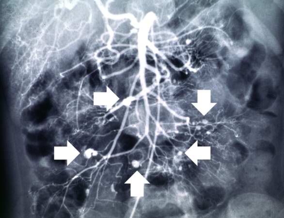
This angiogram of the abdominal viscera demonstrates numerous aneurysms throughout the mesenteric circulation (arrows). |
Polyarteritis nodosa. a case example 2.jpg
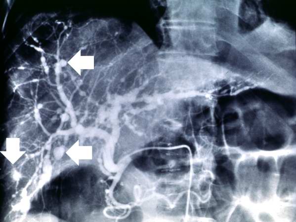
This angiogram of the liver also demonstrates numerous aneurysms throughout the hepatic circulation (arrows). |
Polyarteritis nodosa. a case example 3.jpg
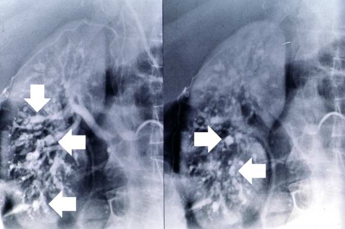
This angiogram of the kidneys demonstrates numerous aneurysmal dilatations in the renal circulation (arrows). |
Polyarteritis nodosa. a case example 4.jpg
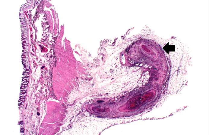
This is a low-power photomicrograph of a mesenteric vessel from this case of polyarteritis nodosa (arrow). The vessel is completely occluded by thrombotic material and the vessel wall is infiltrated with inflammatory cells. |
Polyarteritis nodosa. a case example 5.jpg
This is a higher-power photomicrograph of this mesenteric vessel. Note the thrombotic material occluding the vessel (arrows) and the inflammatory cell infiltrate in the wall of the vessel and in the surrounding adventitia. |
Polyarteritis nodosa. a case example 6.jpg
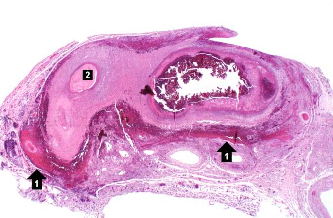
This is another example of a mesenteric artery from this case. There is a marked inflammatory cell response surrounding this vessel, fresh hemorrhage (1), and thrombotic material (2). |
Polyarteritis nodosa. a case example 7.jpg
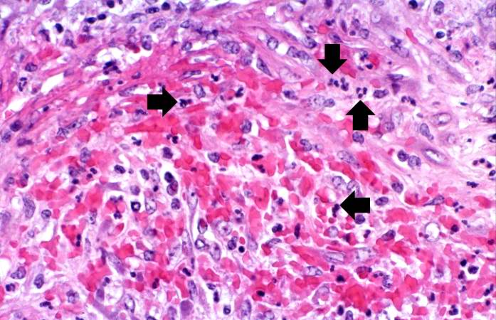
This is a high-power photomicrograph of the vessel wall. There is hemorrhage and infiltration with inflammatory cells--primarily neutrophils (arrows). |
Polyarteritis nodosa. a case example 8.jpg
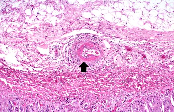
This is a high-power photomicrograph of a small vessel with a rim of fibrinoid necrosis (arrow). |
Polyarteritis nodosa. a case example 9.jpg
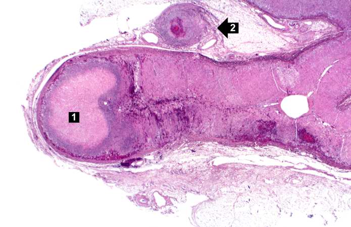
This is a low-power photomicrograph of the adrenal gland. There is an area of necrosis in the adrenal (1) and an affected vessel adjacent to the adrenal (2). |
Polyarteritis nodosa. a case example 10.jpg
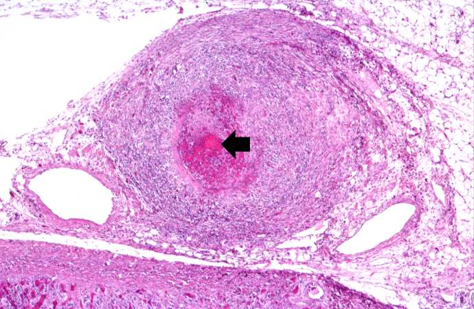
This is a higher-power photomicrograph of the affected vessel from the previous image. The vessel wall is infiltrated with inflammatory cells and the vessel lumen is completely occluded (arrow). |
Polyarteritis nodosa. a case example 11.jpg
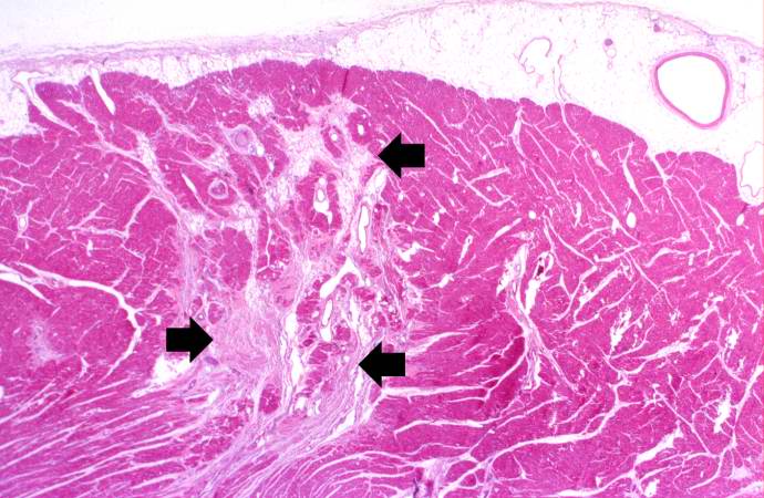
This is a low-power photomicrograph of the heart. There are areas of fibrosis in the myocardium (arrows). Note that the large epicardial coronary artery is normal. |
Polyarteritis nodosa. a case example 12.jpg
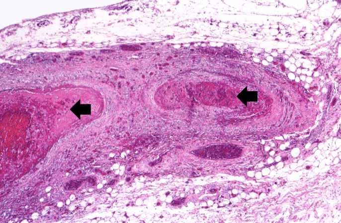
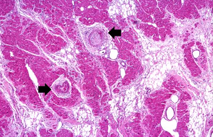
This is a higher-power photomicrograph of the affected vessels in the heart (arrows). There are areas of fibrosis (old infarcts) in the myocardium adjacent to these affected vessels. |
Polyarteritis nodosa. a case example 13.jpg
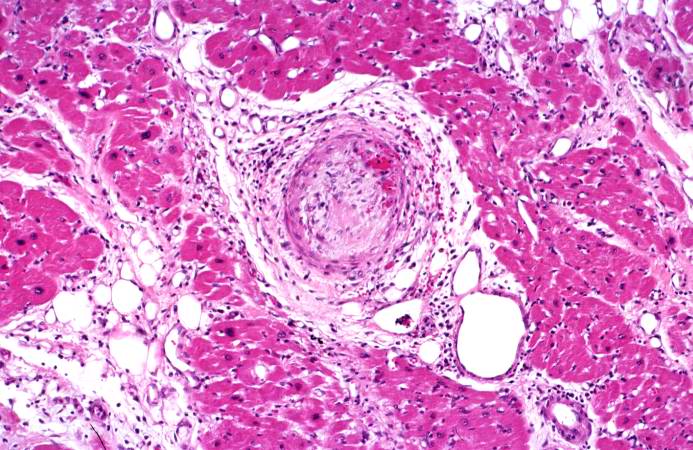
This is a high-power photomicrograph of the affected vessel in the heart. The vessel lumen is completely occluded. |
References
Circulatory system pathology (I, 390-459) | |
|---|---|
| Hypertension | Hypertensive heart disease - Hypertensive nephropathy - Secondary hypertension (Renovascular hypertension) |
| Ischaemic heart disease | Angina pectoris (Prinzmetal's angina) - Myocardial infarction (heart attack) - Dressler's syndrome |
| Pulmonary circulation | Pulmonary embolism - Cor pulmonale |
| Pericardium | Pericarditis - Pericardial effusion - Cardiac tamponade |
| Endocardium/heart valves | Endocarditis - mitral valve (regurgitation, prolapse, stenosis) - aortic valve (stenosis, insufficiency) - pulmonary valve (stenosis, insufficiency) - tricuspid valve (stenosis, insufficiency) |
| Myocardium | Myocarditis - Cardiomyopathy (Dilated cardiomyopathy, Hypertrophic cardiomyopathy, Loeffler endocarditis, Restrictive cardiomyopathy) - Arrhythmogenic right ventricular dysplasia |
| Electrical conduction system of the heart | Heart block: AV block (First degree, Second degree, Third degree) - Bundle branch block (Left, Right) - Bifascicular block - Trifascicular block Pre-excitation syndrome (Wolff-Parkinson-White, Lown-Ganong-Levine) - Long QT syndrome - Adams-Stokes syndrome - Cardiac arrest - Sudden cardiac death Arrhythmia: Paroxysmal tachycardia (Supraventricular, AV nodal reentrant, Ventricular) - Atrial flutter - Atrial fibrillation (Familial) - Ventricular fibrillation - Premature contraction (Atrial, Ventricular) - Ectopic pacemaker - Sick sinus syndrome |
| Other heart conditions | Heart failure - Cardiovascular disease - Cardiomegaly - Ventricular hypertrophy (Left, Right) |
| Cerebrovascular diseases | Stroke - Transient ischemic attack - Intracranial hemorrhage/cerebral hemorrhage: Extra-axial hemorrhage (Epidural hemorrhage, Subdural hemorrhage, Subarachnoid hemorrhage) Intra-axial hematoma (Intraventricular hemorrhages, Intraparenchymal hemorrhage) - Anterior spinal artery syndrome - Binswanger's disease - Moyamoya disease |
| Arteries, arterioles and capillaries | Atherosclerosis (Renal artery stenosis) - Aortic dissection/Aortic aneurysm (Abdominal aortic aneurysm) - Aneurysm - Raynaud's phenomenon/Raynaud's disease - Buerger's disease - Vasculitis/Arteritis (Aortitis) - Intermittent claudication - Arteriovenous fistula - Hereditary hemorrhagic telangiectasia - Spider angioma - Dissection (Carotid artery, Vertebral artery) |
| Veins, lymphatic vessels and lymph nodes | Thrombosis/Phlebitis/Thrombophlebitis (Deep vein thrombosis, May-Thurner syndrome, Portal vein thrombosis, Venous thrombosis, Budd-Chiari syndrome, Renal vein thrombosis, Paget-Schroetter disease) - Varicose veins / Portacaval anastomosis (Hemorrhoid, Esophageal varices, Varicocele, Gastric varices, Caput medusae) - Superior vena cava syndrome - Lymph (Lymphadenitis, Lymphedema, Lymphangitis) |
| Other | Hypotension (Orthostatic hypotension) - Rheumatic fever |
| See also congenital (Q20-Q28, 745-747) | |
Electrocardiography | |
|---|---|
| Overview | History of the EKG • EKG interpretation basics • Normal sinus rhythm |
| EKG Complexes | P wave • QRS complex • ST Segment • T wave • U wave Osborn wave • H wave • K wave • Delta wave |
| EKG Intervals | PR Interval • QRS Interval • ST Interval • QT Interval |
| Conduction System & Bradycardia | Cardiac pacemaker • SA node • AV node• Bundle of His • Purkinje fibers • Sinus bradycardia • First Degree AV Block • Second Degree AV Block • Complete or Third-Degree AV Block • Concealed conduction • AV Junctional Rhythms • LBBB • LAHB • LPHB • RBBB • Trifascicular block |
| Atrial Arrhythmias | Sinus tachycardia • Premature Atrial Contractions (PACs) • Ectopic Atrial Rhythm • Paroxysmal Atrial Tachycardia (PAT) • Paroxysmal Atrial Tachycardia (PAT) with Block • Multifocal Atrial Tachycardia (MAT) • Atrial Flutter • Atrial Fibrillation • Wandering atrial pacemaker |
| Ventricular Arrhythmias | Differential Diagnosis of Tachycardia with a Wide QRS Complex • Accelerated Idioventricular Rhythm • Ventricular Parasystole • Premature Ventricular Contractions (PVCs) • Ventricular tachycardia • Ventricular Fibrillation • Sudden cardiac death |
| EKG Abnormalities in Disease States | Hypertrophy & Dilatation • Right atrial enlargement • Left atrial enlargement • Biatrial enlargement • Left Ventricular Hypertrophy • Right Ventricular Hypertrophy • Biventricular Hypertrophy •
Acute myocardial infarction • NSTEMI • STEMI • Right ventricular myocardial infarction • Atrial infarction Pre-excitation Syndromes • Wolff-Parkinson-White Syndrome • Lown Ganong Levine Syndrome • Mahaim Type Preexcitation Cardiomyopathies • Arrhythmogenic Right Ventricular Dysplasia • Dilated Cardiomyopathy • Hypertrophic Cardiomyopathy Drug Effects on the EKG • Adenosine • β-blockers • Digitalis • Quinidine • Procainamide • Disopyramide • Lidocaine • Tocainide and Mexiletine • Phenytoin • Encainide, Flecainide and Propafenone • Amiodarone • Bretylium • Ca Channel Blockers • Phenothiazines • Tricyclic Antidepressants • Lithium Congenital Heart Disease • Dextrocardia • Atrial Septal Defect • Ventricular Septal Defect • Tetralogy of Fallot • Conjoined Twins or Siamese Twins Electrolyte Disturbances • Hyperkalemia • Hypokalemia • Hypercalcemia • Hypocalcemia • Nonspecific Changes Other Heart Diseases • Pericarditis • Myocarditis • Tamponade • Heart Transplantation • Sick Sinus Syndrome • Long QT Syndrome Inherited Disease • Brugada Syndrome Systemic Diseases • CNS Disease • Cardiac Tumors Heart Transplantation • EKG Changes in patient with Heart Transplantation Exogenous Effects • Hypothermia • Chest Trauma • Insect Bites • Electric Injuries |
| Technical Issues and Potential Errors in Interpretation | Artifacts • Lead Placement Errors • The EKG in a Patient with a Pacemaker |
WikiDoc Research Resources for The heart in polyarteritis nodosa | |
|---|---|
| Articles on The heart in polyarteritis nodosa | Most recent articles on The heart in polyarteritis nodosa • Most cited articles on The heart in polyarteritis nodosa • Review articles on The heart in polyarteritis nodosa • Articles on The heart in polyarteritis nodosa in N Eng J Med, Lancet, BMJ |
| Media (Slides, Video, Images, MP3) on The heart in polyarteritis nodosa | Powerpoint slides on The heart in polyarteritis nodosa • Images of The heart in polyarteritis nodosa • Photos of The heart in polyarteritis nodosa • Podcasts & MP3s on The heart in polyarteritis nodosa • Videos on The heart in polyarteritis nodosa |
| Evidence Based Medicine Regarding The heart in polyarteritis nodosa | Cochrane Collaboration on The heart in polyarteritis nodosa • Bandolier on The heart in polyarteritis nodosa • TRIP on The heart in polyarteritis nodosa |
| Cost Effectiveness of The heart in polyarteritis nodosa | Cost Effectiveness of The heart in polyarteritis nodosa |
| Clinical Trials Involving The heart in polyarteritis nodosa | Ongoing Trials on The heart in polyarteritis nodosa at Clinical Trials.gov • Trial results on The heart in polyarteritis nodosa • Clinical Trials on The heart in polyarteritis nodosa at Google |
| Guidelines / Policies / Government Resources (FDA/CDC) Regarding The heart in polyarteritis nodosa | US National Guidelines Clearinghouse on The heart in polyarteritis nodosa • NICE Guidance on The heart in polyarteritis nodosa • NHS PRODIGY Guidance • FDA on The heart in polyarteritis nodosa • CDC on The heart in polyarteritis nodosa |
| Textbook Information on The heart in polyarteritis nodosa | Books and Textbook Information on The heart in polyarteritis nodosa |
| Pharmacology Resources on The heart in polyarteritis nodosa | Dosing of The heart in polyarteritis nodosa • Drug interactions with The heart in polyarteritis nodosa • Side effects of The heart in polyarteritis nodosa • Allergic reactions to The heart in polyarteritis nodosa • Overdose information on The heart in polyarteritis nodosa • Carcinogenicity information on The heart in polyarteritis nodosa • The heart in polyarteritis nodosa in pregnancy • Pharmacokinetics of The heart in polyarteritis nodosa • |
| Genetics, Pharmacogenomics, and Proteinomics of The heart in polyarteritis nodosa | Genetics of The heart in polyarteritis nodosa • Pharmacogenomics of The heart in polyarteritis nodosa • Proteomics of The heart in polyarteritis nodosa |
| Newstories on The heart in polyarteritis nodosa | The heart in polyarteritis nodosa in the news • Be alerted to news on The heart in polyarteritis nodosa • News trends on The heart in polyarteritis nodosa |
| Commentary on The heart in polyarteritis nodosa | Blogs on The heart in polyarteritis nodosa |
| Patient Resources on The heart in polyarteritis nodosa | Patient resources on The heart in polyarteritis nodosa • Discussion groups on The heart in polyarteritis nodosa • Patient Handouts on The heart in polyarteritis nodosa • Directions to Hospitals Treating The heart in polyarteritis nodosa • Risk calculators and risk factors for The heart in polyarteritis nodosa |
| Healthcare Provider Resources on The heart in polyarteritis nodosa | Symptoms of The heart in polyarteritis nodosa • Causes & Risk Factors for The heart in polyarteritis nodosa • Diagnostic studies for The heart in polyarteritis nodosa • Treatment of The heart in polyarteritis nodosa |
| Continuing Medical Education (CME) Programs on The heart in polyarteritis nodosa | CME Programs on The heart in polyarteritis nodosa |
| International Resources on The heart in polyarteritis nodosa | The heart in polyarteritis nodosa en Espanol • The heart in polyarteritis nodosa en Francais |
| Business Resources on The heart in polyarteritis nodosa | The heart in polyarteritis nodosa in the Marketplace • Patents on The heart in polyarteritis nodosa |
| Informatics Resources on The heart in polyarteritis nodosa | List of terms related to The heart in polyarteritis nodosa |
| ||||
Acknowledgement and Attribution Regarding Sources of Content
Some of the initial content on this page may be incorporated in part from copyleft sources in the public domain including wikis such as Wikipedia and AskDrWiki. Drug information for patients came from the The National Library of Medicine. Infectious disease information may have come from the Centers for Disease Control (CDC). Differential Diagnoses are drawn from clinicians as well as an amalgamation of 3 sources: 1.The Disease Database; 2. Kahan, Scott, Smith, Ellen G. In A Page: Signs and Symptoms. Malden, Massachusetts: Blackwell Publishing, 2004:3; 3. Sailer, Christian, Wasner, Susanne. Differential Diagnosis Pocket. Hermosa Beach, CA: Borm Bruckmeir Publishing LLC, 2002:7 .
