SVG anatomy
|
WikiDoc Resources for SVG anatomy |
|
Articles |
|---|
|
Most recent articles on SVG anatomy Most cited articles on SVG anatomy |
|
Media |
|
Powerpoint slides on SVG anatomy |
|
Evidence Based Medicine |
|
Clinical Trials |
|
Ongoing Trials on SVG anatomy at Clinical Trials.gov Clinical Trials on SVG anatomy at Google
|
|
Guidelines / Policies / Govt |
|
US National Guidelines Clearinghouse on SVG anatomy
|
|
Books |
|
News |
|
Commentary |
|
Definitions |
|
Patient Resources / Community |
|
Patient resources on SVG anatomy Discussion groups on SVG anatomy Patient Handouts on SVG anatomy Directions to Hospitals Treating SVG anatomy Risk calculators and risk factors for SVG anatomy
|
|
Healthcare Provider Resources |
|
Causes & Risk Factors for SVG anatomy |
|
Continuing Medical Education (CME) |
|
International |
|
|
|
Business |
|
Experimental / Informatics |
Editor-In-Chief: C. Michael Gibson, M.S., M.D. [1]
The great saphenous vein (GSV) originates from where the dorsal vein of the first digit (the large toe) merges with the dorsal venous arch of the foot.
After passing anterior to the medial malleolus (where it often can be visualized and palpated), it runs up the medial side of the leg. At the knee, it runs over the posterior border of the medial epicondyle of the femur bone.
The great saphenous vein then courses laterally to lie on the anterior surface of the thigh before entering an opening in the fascia lata called the saphenous opening. It joins with the femoral vein in the region of the femoral triangle at the saphenofemoral junction.
The small saphenous vein (also lesser saphenous vein) is originated where the dorsal vein from the fifth digit (smallest toe) merges with the dorsal venous arch of the foot, which attaches to the great saphenous vein. It is considered a superficial vein and is subcutaneous (just under the skin). From its origin, it courses around the lateral aspect of the foot (inferior and posterior to the lateral malleolus) and runs along the posterior aspect of the leg (with the sural nerve), passes between the heads of the gastrocnemius muscle, and drains into the popliteal vein, approximately at or above the level of the knee joint.
-
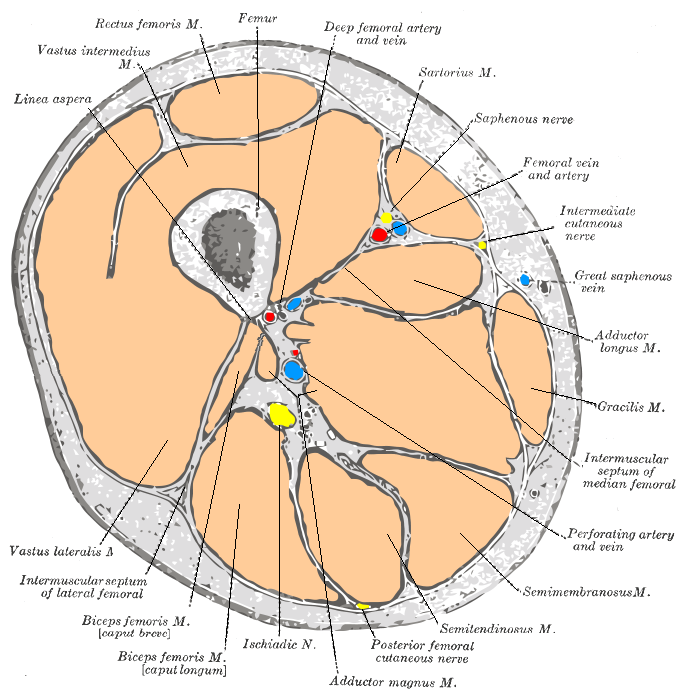
Cross-section through the middle of the thigh.
-
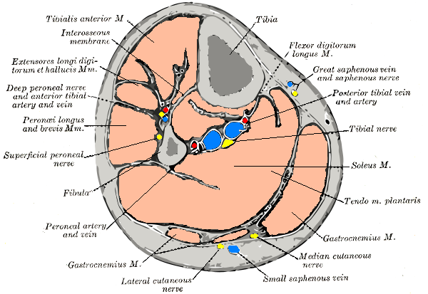
Cross-section through middle of leg.
-
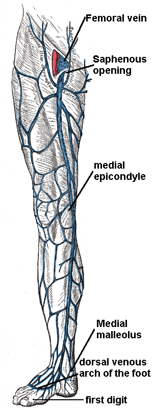
The great saphenous vein and landmarks along its course
-
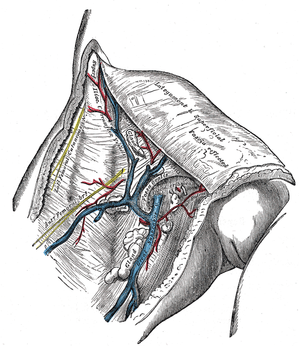
The great saphenous vein and its tributaries at the fossa ovalis in the groin.
-
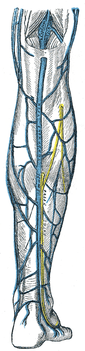
Small saphenous vein and its tributaries.


