Princeps pollicis artery
Editor-In-Chief: C. Michael Gibson, M.S., M.D. [1]
Please Take Over This Page and Apply to be Editor-In-Chief for this topic: There can be one or more than one Editor-In-Chief. You may also apply to be an Associate Editor-In-Chief of one of the subtopics below. Please mail us [2] to indicate your interest in serving either as an Editor-In-Chief of the entire topic or as an Associate Editor-In-Chief for a subtopic. Please be sure to attach your CV and or biographical sketch.
The princeps pollicis (principal artery of the thumb) arises from the radial artery just as it turns medially towards the deep part of the hand; it descends between the first dorsal interosseous muscle and the oblique head of the adductor pollicis, along the medial side of the first metacarpal bone to the base of the proximal phalanx, where it lies beneath the tendon of the flexor pollicis longus muscle and divides into two branches.
These make their appearance between the medial and lateral insertions of the adductor pollicis, and run along the sides of the thumb, forming an arch on the palmar surface of the distal phalanx, from which branches are distributed to the integument and subcutaneous tissue of the thumb.
Additional images
-
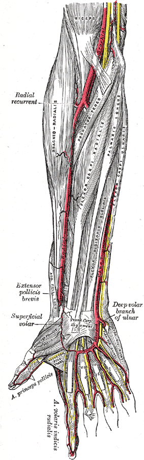 The radial and ulnar arteries. (Arteria princeps pollicis visible at lower left.)
 -
 Ulnar and radial arteries. Deep view.

External links
- Template:UMichAtlas ("Palm of the hand, deep dissection, anterior view")
- Template:UMichAtlas ("Dorsum of the hand, deep dissection, posterior view")
- Template:EMedicineDictionary