Inguinal ligament
Overview
The inguinal ligament is a band running from the pubic tubercle to the anterior superior iliac spine. Its anatomy is very important for operating on hernia patients.
It forms the base of the inguinal canal which is the place from where the inguinal hernia develops.
The inguinal ligament runs from the anterior superior iliac spine of the ilium to the pubic tubercle of the pubic bone. It is formed by the external abdominal oblique aponeurosis and is continuous with the fascia lata of the thigh.
Eponym
It is also referred to as Poupart's ligament, because Poupart gave it its relevance to hernial repair (he called it "le suspenseur de l'abdomen", the suspender of the abdomen). It is less frequently termed the Fallopian ligament.[1][2]
Function
The ligament serves to contain soft tissues as they course anteriorly from the trunk to the lower extremity. This structure demarcates the superior border of the femoral triangle.[3]
Additional images
-
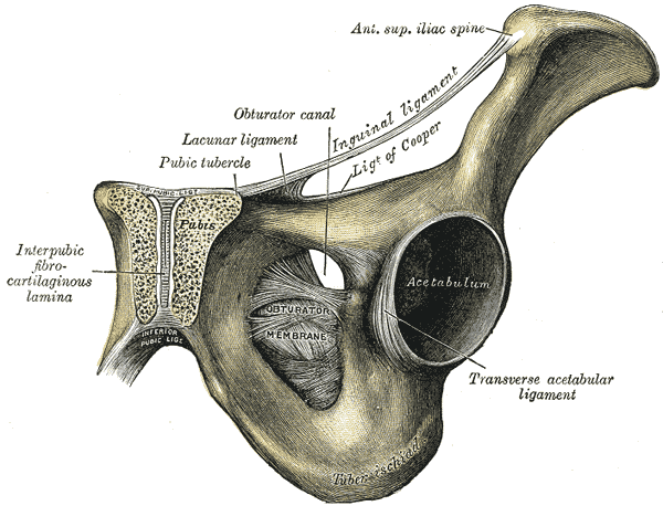
 Articulations of pelvis. Anterior view.
 -
-
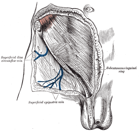 The subcutaneous inguinal ring.
 -
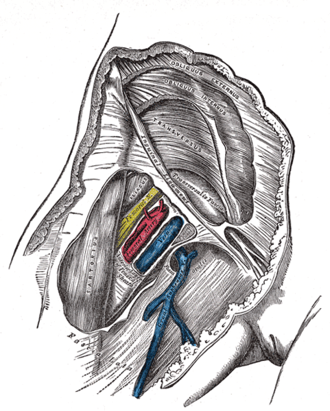 Femoral sheath laid open to show its three compartments.


-
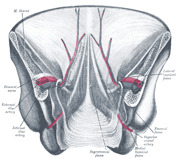
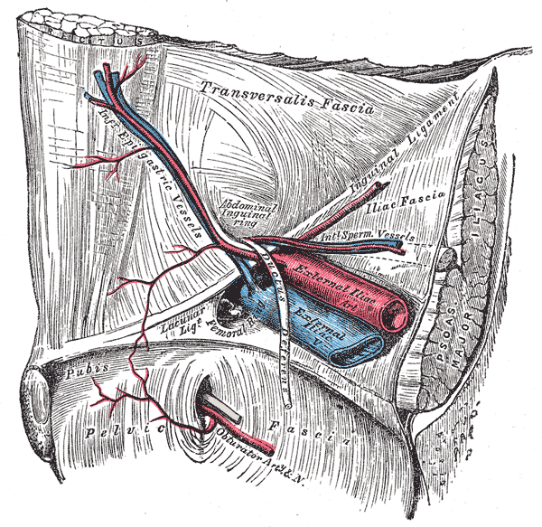 The relations of the femoral and abdominal inguinal rings, seen from within the abdomen. Right side.
 -
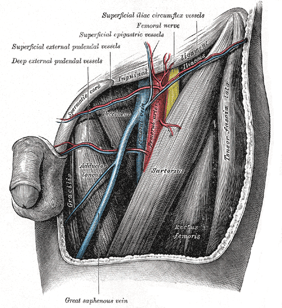 The left femoral triangle.
 -
 Posterior view of the anterior abdominal wall in its lower half. The peritoneum is in place, and the various cords are shining through.


References
- ↑ Template:WhoNamedIt
- ↑ F. Poupart. Chirurgie complète. Paris, 1695.
- ↑ Ryan, Jeffrey M.; Starkey, Chad (2002). Evaluation of orthopedic and athletic injuries. Philadelphia: F.A. Davis Co. ISBN 0-8036-0791-1.
External links
- Template:GPnotebook
- Template:SUNYAnatomyFigs - "Deep muscles of the anterior thigh."
- Template:SUNYAnatomyLabs - "Anterior Abdominal Wall: Osteology and Surface Anatomy "
- Template:SUNYAnatomyLabs - "Anterior Abdominal Wall: The Inguinal Ligament"
- Template:SUNYAnatomyImage
- Template:SUNYAnatomyImage
- Diagram at gensurg.co.uk