Optic chiasm
Overview
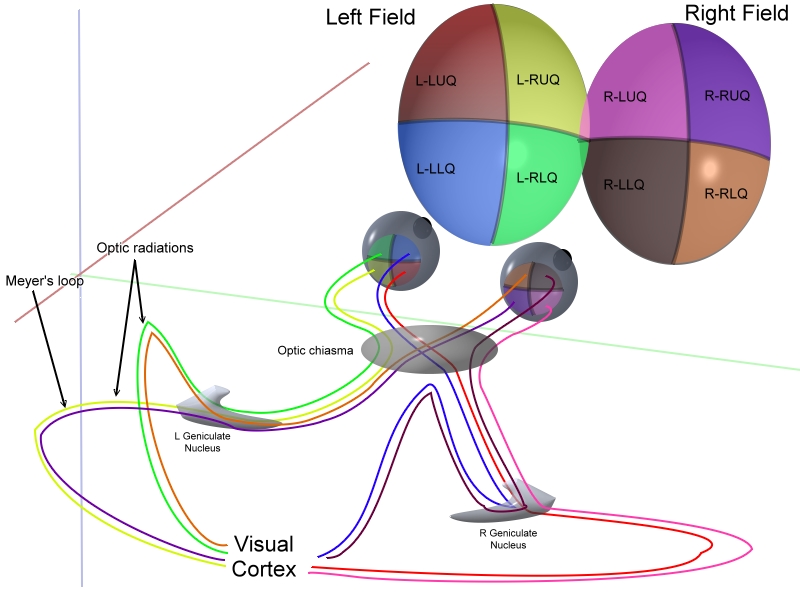
The optic chiasm (Greek χίασμα, "crossing", from the Greek χλαζειν 'to mark with an X', after the Greek letter 'Χ', chi) is the part of the brain where the optic nerves partially cross.
Pathways
Specifically, in the optic chiasm, the nerves connected to the right eye that attend to the right temporal visual field (located in the nasal portion of the right retina) cross to the left half of the brain, while the nerves from the left eye that attend to the left temporal visual field (located in the nasal portion of the left retina) cross to the right half of the brain.
This allows for parts of both eyes that attend to the right visual field to be processed in the left visual system in the brain, and vice versa.
Optic chiasm in cats
In Siamese cats with certain genotypes of the albino gene, this wiring is disrupted, with less of the nerve-crossing than is normal, as a number of scholars have reported. [1] To compensate for lack of crossing in their brains, they cross their eyes (strabismus). [2]
This is also seen in albino tigers, as Guillery & Kaas report.[3]
Additional images
-
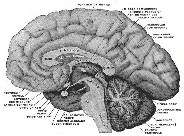
Mesal aspect of a brain sectioned in the median sagittal plane.
-

Median sagittal section of brain.
-
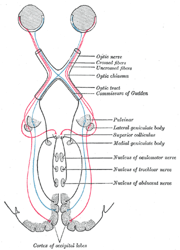
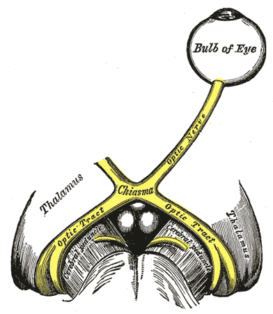
Scheme showing central connections of the optic nerves and optic tracts.
-
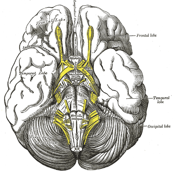
Base of brain.
-

Coronal section of brain through anterior commissure.
-
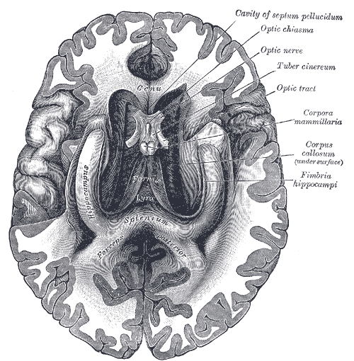
The fornix and corpus callosum from below.
-
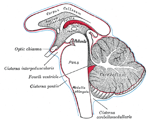
Diagram showing the positions of the three principal subarachnoid cisternæ.
-
The left optic nerve and the optic tracts.
-
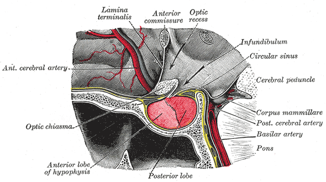
The hypophysis cerebri in position. Shown in sagittal section.
-
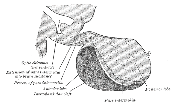
Median sagittal through the hypophysis of an adult monkey. Semidiagrammatic.
-
3D schematic representation of optic tracts


References
- Jeffery, Glen. Architecture of the Optic Chiasm and the Mechanisms That Sculpt Its Development Physiol Rev, Oct 2001; 81: 1393 - 1414.
External links
Template:Visual system Template:Diencephalon