Macula
Template:Infobox Anatomy Editor-In-Chief: C. Michael Gibson, M.S., M.D. [1]
The macula or macula lutea (from Latin macula, "spot" + lutea, "yellow") is an oval yellow spot near the center of the retina of the human eye. It has a diameter of about 1.5 mm and is often histologically defined as having two or more layers of ganglion cells. Near its center is the fovea, a small pit that contains the largest concentration of cone cells in the eye and is responsible for central vision.
It is specialized for high acuity vision. Within the macula are the fovea and foveola which contain a high density of cones (photoreceptors with high acuity).
Clinical significance
Whereas loss of peripheral vision may go unnoticed for some time, damage to the macula will result in loss of central vision, which is usually immediately obvious. The progressive destruction of the macula is a disease known as macular degeneration and leads to the creation of a macular hole. Macular holes can rarely be caused by trauma, if a severe blow is delivered perfectly it can burst the blood vessels going to the macula, destroying it.
Visual input to the macula occupies a substantial portion of the brain's visual capacity. As a result, some forms of visual field loss can occur without involving the macula; this is termed macular sparing. (For example, visual field testing might demonstrate homonymous hemianopsia with macular sparing.) This finding can be very informative for the ophthalmologist.
See also
Additional images
-
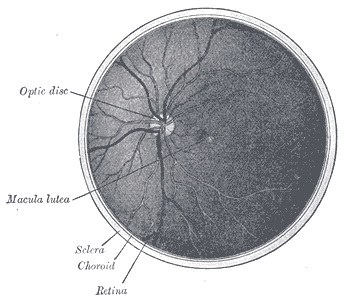 The interior of the posterior half of the left eyeball.

Template:Eye
Template:Visual system
de:Gelber Fleck (Auge) el:Ωχρή κηλίδα lt:Geltonoji dėmė nl:Gele vlek no:Macula lutea simple:Yellow spot sv:Gula fläcken