Gluteus medius muscle
Overview
The gluteus medius, one of the three gluteal muscles, is a broad, thick, radiating muscle, situated on the outer surface of the pelvis.
Its posterior third is covered by the gluteus maximus, its anterior two-thirds by the gluteal aponeurosis, which separates it from the superficial fascia and integument.
Origin and insertion
It arises from the outer surface of the ilium between the iliac crest and posterior gluteal line above, and the anterior gluteal line below; it also arises from the gluteal aponeurosis covering its outer surface.
The fibers converge to a strong flattened tendon, which is inserted into the oblique ridge which runs downward and forward on the lateral surface of the greater trochanter.
Relations
A bursa separates the tendon of the muscle from the surface of the trochanter over which it glides.
Action
The Glutæi medius and minimus abduct the thigh, when the limb is extended, and are principally called into action in supporting the body on one limb, in conjunction with the Tensor fasciæ latæ.
Their anterior fibers, by drawing the greater trochanter forward, rotate the thigh inward, in which action they are also assisted by the Tensor fasciæ latæ. When the hip is flexed to ninety degrees however the glutæi medius aids in rotating the thigh outwards.
Variations
The posterior border may be more or less closely united to the piriformis, or some of the fibers end on its tendon.
The posterior fibres of gluteus medius contract to produce hip extension, lateral rotation and abduction. During gait, the posterior fibres help to decelerate internal rotation of the femur at the end of swing phase.
Additional images
-
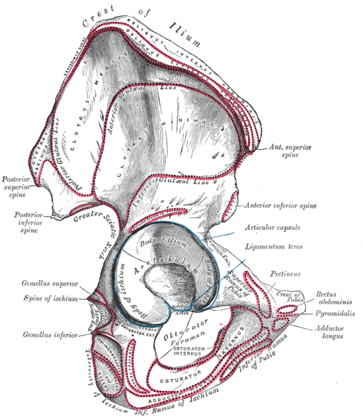 Right hip bone. External surface.
 -
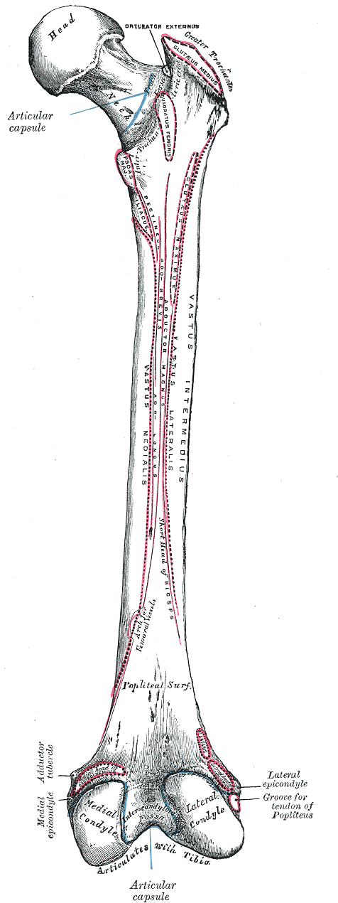 Right femur. Posterior surface.
 -
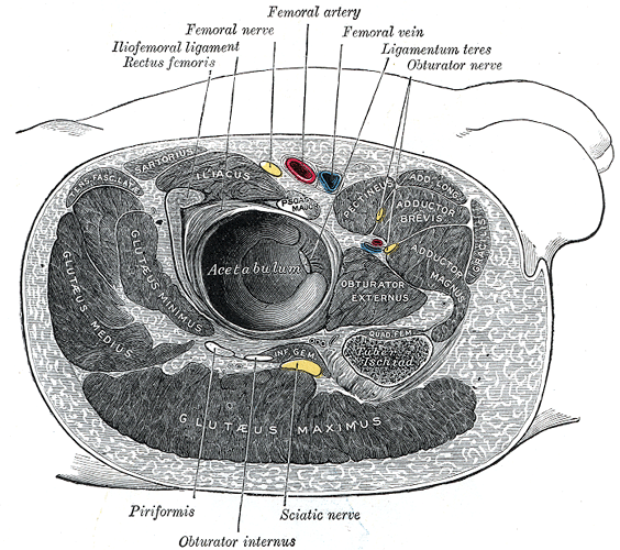 Structures surrounding right hip-joint.
 -
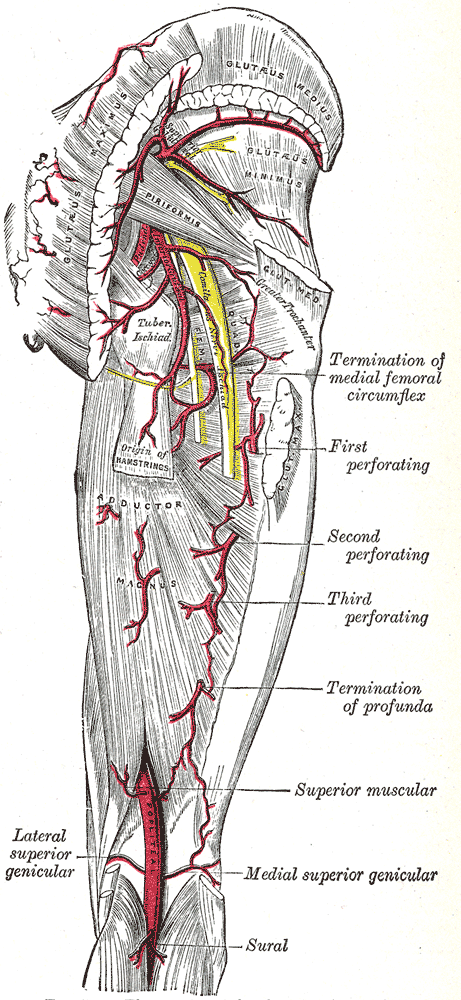 The arteries of the gluteal and posterior femoral regions.
 -
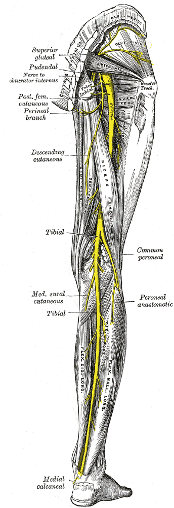 Nerves of the right lower extremity Posterior view.
 -
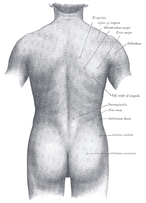 Surface anatomy of the back.


See also
External links
Template:Gray's Template:Muscles of lower limb
cs:Střední sval hýžďový de:Musculus gluteus medius he:שריר העכוז האמצעי it:Medio gluteo la:Musculus gluteus medius fi:Keskimmäinen pakaralihas Template:WH Template:WikiDoc Sources