Frontal sinus
|
WikiDoc Resources for Frontal sinus |
|
Articles |
|---|
|
Most recent articles on Frontal sinus Most cited articles on Frontal sinus |
|
Media |
|
Powerpoint slides on Frontal sinus |
|
Evidence Based Medicine |
|
Clinical Trials |
|
Ongoing Trials on Frontal sinus at Clinical Trials.gov Trial results on Frontal sinus Clinical Trials on Frontal sinus at Google
|
|
Guidelines / Policies / Govt |
|
US National Guidelines Clearinghouse on Frontal sinus NICE Guidance on Frontal sinus
|
|
Books |
|
News |
|
Commentary |
|
Definitions |
|
Patient Resources / Community |
|
Patient resources on Frontal sinus Discussion groups on Frontal sinus Patient Handouts on Frontal sinus Directions to Hospitals Treating Frontal sinus Risk calculators and risk factors for Frontal sinus
|
|
Healthcare Provider Resources |
|
Causes & Risk Factors for Frontal sinus |
|
Continuing Medical Education (CME) |
|
International |
|
|
|
Business |
|
Experimental / Informatics |
The frontal sinuses, situated behind the superciliary arches, are rarely symmetrical, and the septum between them frequently deviates to one or other side of the middle line.
Their average measurements are as follows: height, 3 cm.; breadth, 2.5 cm.; depth from before backward, 2.5 cm.
Each opens into the anterior part of the corresponding middle meatus of the nose through the frontonasal duct which traverses the anterior part of the labyrinth of the ethmoid. These structures then open into the hiatus semilunaris in the middle meatus.
The mucuous membrane in this sinus is innervated by the supraorbital nerve and supplied by the supraorbital artery and anterior ethmoidal artery.
Absent at birth, they are generally fairly well developed between the seventh and eighth years, but only reach their full size after puberty.
Additional images
-
-

Frontal bone. Inner surface.
-

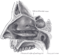
Medial wall of left orbit.
-
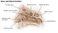
Lateral wall of nasal cavity.
-
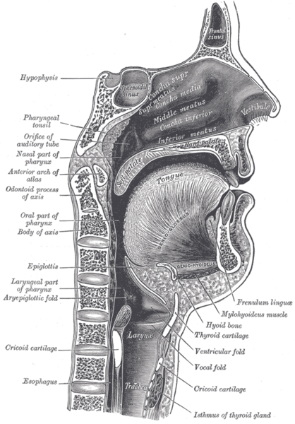
Sagittal section of nose mouth, pharynx, and larynx.
-
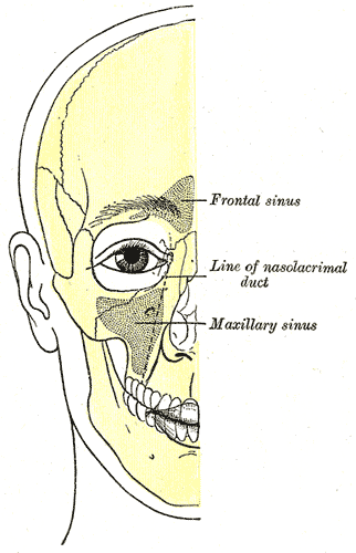
Outline of bones of face, showing position of air sinuses.


See also
External links
- Template:SUNYAnatomyLabs
- Template:NormanAnatomy (Template:NormanAnatomyFig, Template:NormanAnatomyFig)
Template:Gray's Template:Skull