File:Subependymal giant cell astrocytoma MRI 3.jpg
Jump to navigation
Jump to search
No higher resolution available.
Subependymal_giant_cell_astrocytoma_MRI_3.jpg (387 × 512 pixels, file size: 121 KB, MIME type: image/jpeg)
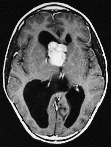
Subependymal giant cell astrocytoma of the type associated with tuberous sclerosis are typically bulky, contrast-enhancing masses in the region of the foramen of Monro. Most overlie the head of the caudate nucleus. Foramen obstruction has produced hydrocephalus.
File history
Click on a date/time to view the file as it appeared at that time.
| Date/Time | Thumbnail | Dimensions | User | Comment | |
|---|---|---|---|---|---|
| current | 00:24, 3 November 2015 | | 387 × 512 (121 KB) | Sujit Routray (talk | contribs) | Subependymal giant cell astrocytoma of the type associated with tuberous sclerosis are typically bulky, contrast-enhancing masses in the region of the foramen of Monro. Most overlie the head of the caudate nucleus. Foramen obstruction has produced hydr... |
You cannot overwrite this file.
File usage
The following file is a duplicate of this file (more details):
There are no pages that use this file.