File:Pilocytic astrocytoma micro 4.jpeg
Jump to navigation
Jump to search
Size of this preview: 800 × 579 pixels. Other resolution: 1,017 × 736 pixels.
Original file (1,017 × 736 pixels, file size: 154 KB, MIME type: image/jpeg)
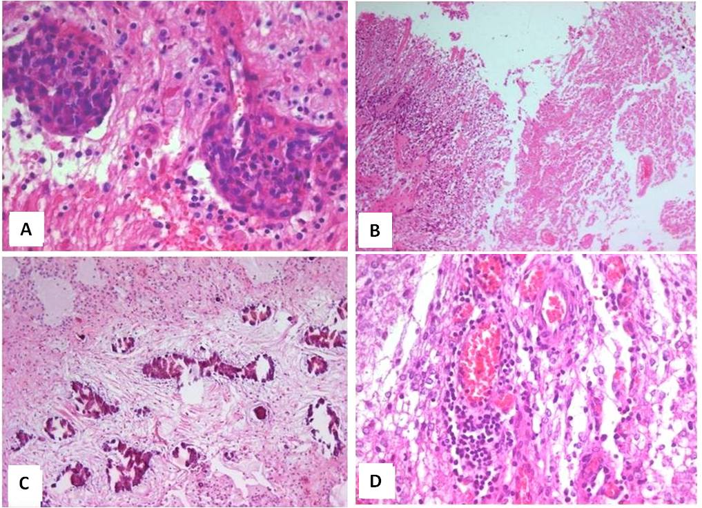
Some other histopathological aspects of pilocytic astrocytoma at the H&E stain. A- Microvascular proliferation (200x) B- Areas of necrosis may be seen in some tumors – in this case, the necrosis is at right (40x). C- Calcification foci near a microcystic area of tumor (40x). D- Lymphocytic cuffs may be present (200x).
File history
Click on a date/time to view the file as it appeared at that time.
| Date/Time | Thumbnail | Dimensions | User | Comment | |
|---|---|---|---|---|---|
| current | 22:22, 26 October 2015 | | 1,017 × 736 (154 KB) | Sujit Routray (talk | contribs) | Some other histopathological aspects of pilocytic astrocytoma at the H&E stain. A- Microvascular proliferation (200x) B- Areas of necrosis may be seen in some tumors – in this case, the necrosis is at right (40x). C- Calcification foci near a microcy... |
| 22:21, 26 October 2015 |  | 1,017 × 736 (154 KB) | Sujit Routray (talk | contribs) | Some other histopathological aspects of pilocytic astrocytoma at the H&E stain. A- Microvascular proliferation (200x) B- Areas of necrosis may be seen in some tumors – in this case, the necrosis is at right (40x). C- Calcification foci near a microcy... |
You cannot overwrite this file.
File usage
There are no pages that use this file.