File:Pilocytic astrocytoma micro 2.png
Jump to navigation
Jump to search
Size of this preview: 800 × 600 pixels. Other resolution: 1,144 × 858 pixels.
Original file (1,144 × 858 pixels, file size: 1.21 MB, MIME type: image/png)
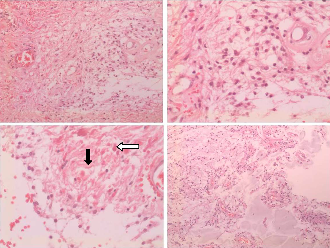
Histopathological aspects of pilocytic astrocytoma. All microphotographs represent H&E stain. A- Biphasic pattern – high cellularity at left and lower cellularity at bottom right (100x). B- Round and piloid cells constitute this tumor. Note the vasculature, with typical endothelia (200x). C- Rosenthal fibers (black arrow) and eosinophilic granular body (white arrow) can be seen in higher cellular areas (200x). D- At bottom, a microcystic area is seen (40x).
File history
Click on a date/time to view the file as it appeared at that time.
| Date/Time | Thumbnail | Dimensions | User | Comment | |
|---|---|---|---|---|---|
| current | 22:14, 26 October 2015 | | 1,144 × 858 (1.21 MB) | Sujit Routray (talk | contribs) | Histopathological aspects of pilocytic astrocytoma. All microphotographs represent H&E stain. A- Biphasic pattern – high cellularity at left and lower cellularity at bottom right (100x). B- Round and piloid cells constitute this tumor. Note the vascu... |
| 22:12, 26 October 2015 |  | 1,144 × 858 (1.21 MB) | Sujit Routray (talk | contribs) | Histopathological aspects of pilocytic astrocytoma. All microphotographs represent H&E stain. A- Biphasic pattern – high cellularity at left and lower cellularity at bottom right (100x). B- Round and piloid cells constitute this tumor. Note the vascu... |
You cannot overwrite this file.
File usage
There are no pages that use this file.