File:Pilocytic astrocytoma MRI original 2.PNG
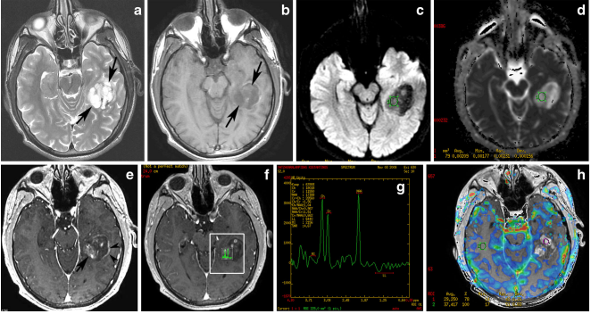
Hemispheric pilocytic astrocytoma in a 26-year-old man. a An axial T2-weighted image demonstrates a temporally located, lobular mass that is very hyperintense (arrows) without vasogenic edema. b On this T1- weighted image, the lesion is hypointense compared with grey matter (arrows). c The lesion exhibits low signal on diffusion-weighted imaging, with high signal intensity on an ADC map (d). e On this post-contrast T1-weighted image, nodular (arrow) and peripheral rim enhancements (arrowheads) are visible. f, g MR spectroscopy with TE 144 ms revealed an elevated choline peak without reduction of the NAA peak. h Perfusion MRI reveals low rCBV in the solid (enhanced) part of the tumor.
File history
Click on a date/time to view the file as it appeared at that time.
| Date/Time | Thumbnail | Dimensions | User | Comment | |
|---|---|---|---|---|---|
| current | 15:34, 3 November 2015 | | 662 × 352 (288 KB) | Sujit Routray (talk | contribs) | Hemispheric pilocytic astrocytoma in a 26-year-old man. '''a''' An axial T2-weighted image demonstrates a temporally located, lobular mass that is very hyperintense (arrows) without vasogenic edema. '''b''' On this T1- weighted image, the lesion is hyp... |
| 15:33, 3 November 2015 |  | 662 × 352 (288 KB) | Sujit Routray (talk | contribs) | Hemispheric pilocytic astrocytoma in a 26-year-old man. '''a''' An axial T2-weighted image demonstrates a temporally located, lobular mass that is very hyperintense (arrows) without vasogenic edema. '''b''' On this T1- weighted image, the lesion is hyp... |
You cannot overwrite this file.
File usage
There are no pages that use this file.