File:Pilocytic astrocytoma MRI original 1.PNG
Jump to navigation
Jump to search
No higher resolution available.
Pilocytic_astrocytoma_MRI_original_1.PNG (575 × 433 pixels, file size: 229 KB, MIME type: image/png)
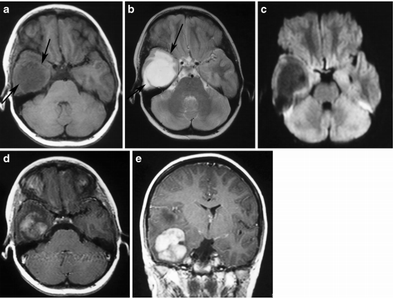
Supratentorial pilocytic astrocytoma with solid enhancement and vasogenic edema. a An axial T1-weighted image shows a wellcircumscribed, hypointense cerebral mass (arrows). b An axial T2- weighted image shows a hyperintense mass. Note the vasogenic edema around the mass (arrows). c The lesion exhibits low signal on diffusion weighted imaging. Contrast-enhanced axial (d) and (e) T1-weighted images demonstrate inhomogeneous enhancement of the mass.
File history
Click on a date/time to view the file as it appeared at that time.
| Date/Time | Thumbnail | Dimensions | User | Comment | |
|---|---|---|---|---|---|
| current | 15:27, 3 November 2015 | | 575 × 433 (229 KB) | Sujit Routray (talk | contribs) | Supratentorial pilocytic astrocytoma with solid enhancement and vasogenic edema. '''a''' An axial T1-weighted image shows a wellcircumscribed, hypointense cerebral mass (arrows). '''b''' An axial T2- weighted image shows a hyperintense mass. Note the v... |
| 15:27, 3 November 2015 |  | 575 × 433 (229 KB) | Sujit Routray (talk | contribs) | Supratentorial pilocytic astrocytoma with solid enhancement and vasogenic edema. '''a''' An axial T1-weighted image shows a wellcircumscribed, hypointense cerebral mass (arrows). '''b''' An axial T2- weighted image shows a hyperintense mass. Note the v... |
You cannot overwrite this file.
File usage
There are no pages that use this file.