File:Oligodendroglioma MRI axial T2.jpg
Jump to navigation
Jump to search
Size of this preview: 549 × 599 pixels. Other resolution: 577 × 630 pixels.
Original file (577 × 630 pixels, file size: 32 KB, MIME type: image/jpeg)
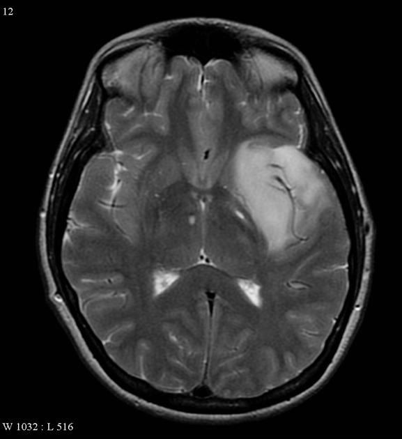
A relatively well circumscribed mass involves the temporal lobe and insular cortex, without convincing enhancement, and minimal restricted diffusion.
File history
Click on a date/time to view the file as it appeared at that time.
| Date/Time | Thumbnail | Dimensions | User | Comment | |
|---|---|---|---|---|---|
| current | 14:51, 8 October 2015 | | 577 × 630 (32 KB) | Sujit Routray (talk | contribs) | A relatively well circumscribed mass involving the temporal lobe and insular cortex, without convincing enhancement, and minimal restricted diffusion. |
| 14:50, 8 October 2015 |  | 577 × 630 (32 KB) | Sujit Routray (talk | contribs) | A relatively well circumscribed mass involves the temporal lobe and insular cortex, without convincing enhancement, and minimal restricted diffusion. |
You cannot overwrite this file.
File usage
There are no pages that use this file.