File:N1.jpg
Jump to navigation
Jump to search

Size of this preview: 789 × 599 pixels. Other resolution: 1,132 × 860 pixels.
Original file (1,132 × 860 pixels, file size: 156 KB, MIME type: image/jpeg)
Summary
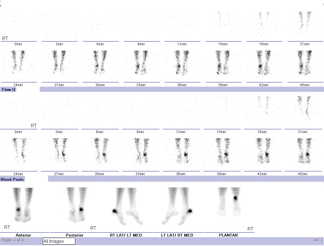
Dynamic flow and blood pool images show increased perfusion and vascularity at the right heel. Delayed static images show an intense increase in tracer uptake localized to the posterior aspect of the right calcaneum highly suspicious of a fracture.
Case courtesy of Assoc Prof Craig Hacking, <a href="https://radiopaedia.org/">Radiopaedia.org</a>. From the case <a href="https://radiopaedia.org/cases/34978">rID: 34978</a>
File history
Click on a date/time to view the file as it appeared at that time.
| Date/Time | Thumbnail | Dimensions | User | Comment | |
|---|---|---|---|---|---|
| current | 18:56, 8 March 2020 |  | 1,132 × 860 (156 KB) | DrMars (talk | contribs) | Dynamic flow and blood pool images show increased perfusion and vascularity at the right heel. Delayed static images show an intense increase in tracer uptake localized to the posterior aspect of the right calcaneum highly suspicious of a fracture.... |
You cannot overwrite this file.
File usage
The following 4 pages use this file: